
Figures & data
Figure 1 Chemical structure of donepezil (CAS: 120014-06-4).
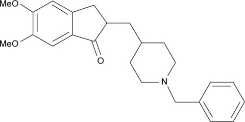
Figure 2 SEM micrographs of the prepared liposomes of DNP.
Abbreviations: SEM, scanning electron microscopy; DNP, donepezil.
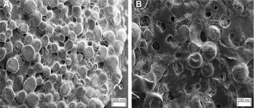
Figure 3 In vitro release of DNP from liposomes (mean ± standard deviation, n=3).
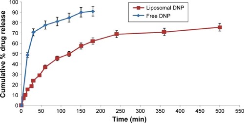
Table 1 Encapsulation efficiency and vesicle size of the prepared liposomes during storage at 4°C and 25°C over a period of 3 months
Table 2 Pharmacokinetic parameters of DNP in plasma post various routes of administration (mean ± SD, n=6)
Figure 4 Mean plasma concentration and time profile of donepezil with various routes of administration (mean ± standard deviation, n=6).
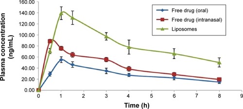
Table 3 Pharmacokinetic parameters of DNP in brain post various routes of administration (mean ± SD, n=6)
Figure 5 Mean brain concentrations and time profile of donepezil with various routes of administration (mean ± standard deviation, n=6).
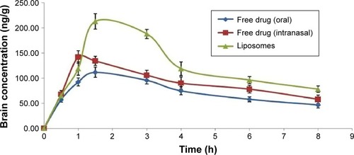
Figure 6 Histopathological examinations of heart, lung, kidney, spleen, and liver.
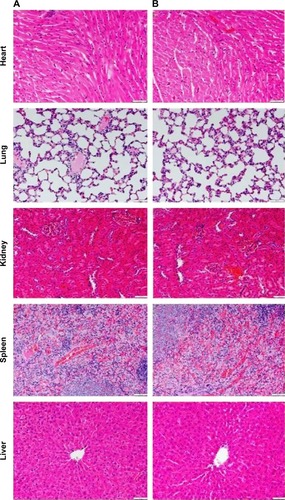
Figure 7 Histopathological examinations of brain and olfactory bulb.
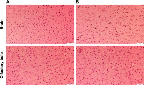
Table 4 Dissolution kinetic parameters for the DNP release from liposomal formulation