Figures & data
Figure 1 Antiproliferation effect of Andro on Jurkat cells.
Notes: Jurkat cells were cultured in 96-well, flat bottom microtiter plates at a density of 1.0×10⁴ cells per well for 24 hours. Then, the cells were treated with different concentrations of Andro for 24, 48, and 72 hours, and then cell viability was determined by MTT assay. Mean ± SD, n=3.
Abbreviations: Andro, Andrographolide; SD, standard deviation; MTT, 3-(4,5-dimethyl-2-thiazolyl)-2,5-diphenyl-2-H-tetrazolium bromide.
Abbreviations: Andro, Andrographolide; SD, standard deviation; MTT, 3-(4,5-dimethyl-2-thiazolyl)-2,5-diphenyl-2-H-tetrazolium bromide.
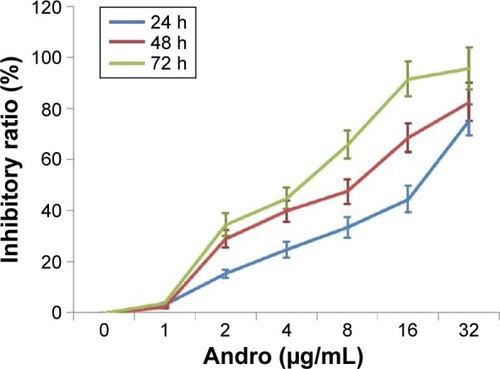
Figure 2 Andro induced apoptosis in Jurkat cells.
Notes: (A and B) Jurkat cells were treated with different concentrations of Andro and/or z-VAD for 48 hours. Cells were digested to form a single-cell suspension with EDTA-free trypsin and stained according to the manufacturer’s instruction provided with the Annexin V-FITC/PI Apoptosis Detection kit. After that, stained cells were analyzed by flow cytometry. (C) Jurkat cells cultured in the media were observed under inverted microscope. (D) Jurkat cells were washed with 1× PBS and harvested on ice. Lysates were prepared using a lysis buffer and then subjected to Western blot analysis to detect capase-3 protein expression. Mean ± SD, n=3. *P<0.05, **P<0.01 versus DMSO group.
Abbreviations: Andro, Andrographolide; EDTA, ethylenediaminetetraacetic acid; FITC, fluorescein isothiocyanate; PI, propidium iodide; DMSO, dimethyl sulfoxide; SD, standard deviation; PBS, phosphate-buffered saline.
Abbreviations: Andro, Andrographolide; EDTA, ethylenediaminetetraacetic acid; FITC, fluorescein isothiocyanate; PI, propidium iodide; DMSO, dimethyl sulfoxide; SD, standard deviation; PBS, phosphate-buffered saline.
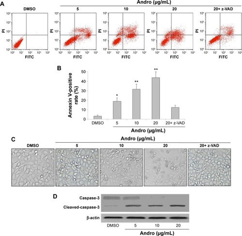
Figure 3 Andro could not induce autophagy in Jurkat cells.
Notes: (A) Jurkat cells were treated with different concentrations of Andro and subjected to quantitative analysis, detecting a positive ratio of MDC staining by flow cytometry. (B) Jurkat cells were washed with 1× PBS and harvested on ice. Lysates were prepared in a lysis buffer, and then subjected to Western blot analysis to detect LC3 protein expression. Mean ± SD, n=3.
Abbreviations: MDC, monodansylcadaverine; DMSO, dimethyl sulfoxide; Andro, Andrographolide; PBS, phosphate-buffered saline; SD, standard deviation.
Abbreviations: MDC, monodansylcadaverine; DMSO, dimethyl sulfoxide; Andro, Andrographolide; PBS, phosphate-buffered saline; SD, standard deviation.
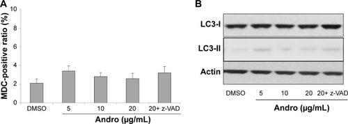
Figure 4 The effects of Andro on PI3K/AKT and MAPK pathways in Jurkat cells.
Notes: (A) Jurkat cells were treated with different concentrations of Andro for 48 hours. Later, the cells were washed with 1× PBS and harvested on ice. Lysates were prepared in a lysis buffer, and then subjected to Western blot analysis to detect p-AKT, p-JNK, p-ERK, p-p38, and p-p53 proteins expression. (B) The cells were harvested and RNA was extracted from cell lysates using Qiagen RNA easy kits; RT-PCR was applied to detect AKT, p38, JNK, ERK RNA level. The target genes’ expression levels were normalized to negative control GAPHD. Mean ± SD, n=3. (C and D) The cells were pretreated with PD98059, SP600125, or SB203580 for 0.5 hours, and then cultured with 20 μg/mL Andro for 48 hours. Later, the cells were harvested for MTT and Annexin V-FITC/PI double staining. *P<0.05, **P<0.01 versus DMSO group.
Abbreviations: Andro, Andrographolide; DMSO, dimethyl sulfoxide; PBS, phosphate-buffered saline; RT-PCR, real-time polymerase chain reaction; FITC, fluorescein isothiocyanate; PI, propidium iodide; SD, standard deviation; MTT, 3-(4,5-dimethyl-2-thiazolyl)-2,5-diphenyl-2-H-tetrazolium bromide.
Abbreviations: Andro, Andrographolide; DMSO, dimethyl sulfoxide; PBS, phosphate-buffered saline; RT-PCR, real-time polymerase chain reaction; FITC, fluorescein isothiocyanate; PI, propidium iodide; SD, standard deviation; MTT, 3-(4,5-dimethyl-2-thiazolyl)-2,5-diphenyl-2-H-tetrazolium bromide.
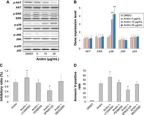
Figure 5 Antitumor effect of Andro on subcutaneous Jurkat xenografts.
Notes: (A) Nude mice bearing Jurkat tumors were randomly separated into four groups: vehicle, 50, 100, and 200 mg/kg Andro. Tumor volumes of mice were monitored twice a week. (B and C) The mice were subjected to euthanasia and the tumors were collected and weighed after 3 weeks’ treatment. *P<0.05 versus Andro group, **P<0.01 versus vehicle group.
Abbreviations: Andro, Andrographolide; SD, standard deviation.
Abbreviations: Andro, Andrographolide; SD, standard deviation.
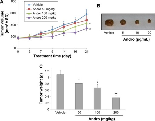
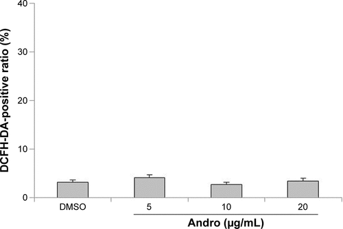
Figure S1 Andro could not induce ROS generation in Jurkat cells.
Notes: Jurkat cells were treated with different concentrations of Andro and subjected to quantitative analysis, detecting a positive ratio of DCFH-DA staining by flow-cytometry. DCFH-DA is a specific marker for ROS detection.
Abbreviations: DCFH-DA, 2′,7′-dichlorofluorescin diacetate; DMSO, dimethyl sulfoxide; Andro, Andrographolide; ROS, reactive oxygen species.
Abbreviations: DCFH-DA, 2′,7′-dichlorofluorescin diacetate; DMSO, dimethyl sulfoxide; Andro, Andrographolide; ROS, reactive oxygen species.