Figures & data
Figure 1 Timeline for streptozotocin (STZ) injection, exosome transplantation and metabolic analyses.
Notes: (A) High-dose of STZ (200mg/kg, once) was administered in mice to build a diabetic model. Exosomes derived from MIN6 cells or PBS were transplanted in situ pancreas after non-fasting glucose levels reached 11.1mmol/l. Body weight and non-fasted blood glucose concentrations were monitored once every week. (B) Low dose of STZ (140mg/kg, once) was intraperitoneally administered in mice. An intraperitoneal glucose tolerance test (IPGTT) was performed on day 7, day 17 and day 35. Exosomes or PBS were transplanted in situ pancreas on day 10 and were given via tail vein injection on day 21.
Abbreviations: IPGTT, intraperitoneal glucose tolerance test; STZ, streptozotocin.
Abbreviations: IPGTT, intraperitoneal glucose tolerance test; STZ, streptozotocin.
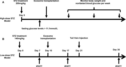
Figure 2 Characterization of exosomes derived from MIN6 cells.
Notes: (A) Size distribution of exosomes evaluated by a ZetaSizer Nano ZS. (B) Transmission electron microscopy image of the exosomes, scale bar: 100nm. (C) Western blot analysis of exosome marker CD9 and CD63. (D) Cell viability in INS-1(832/13) (INS-1) cells. INS-1 cells were exposed to palmitate (PA, 0.4 mmol/L) and different concentrations of exosomes (Exo, 0–1.5-4.5–9.0X10Citation9/mL) for 48 hrs. Data were presented as fold-change of INS-1 cell viability over cells treated without palmitate and exosomes. * P<0.05, ** P<0.01, between indicated groups (n=4).
Abbreviations: PA, palmitate; Exo, exosomes.
Abbreviations: PA, palmitate; Exo, exosomes.
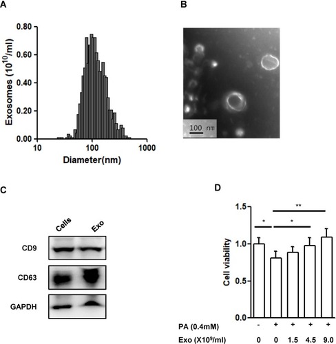
Figure 3 Effect of exosomes (Exo) on streptozotocin (STZ)-induced diabetic mice.
Notes: Male C57 mice were administrated with high-dose of STZ (200mg/kg), and Exo was transplanted afterwards. (A) Non-fasting blood glucose levels, (B) survival time, (C) body weight, (D) serum insulin and (E) insulin content extract from pancreas were compared. Data were showed as mean ± SEM. *P<0.05, **P<0.01, ***P<0.001, between indicated groups (Control, n=3; STZ, n=5; STZ+Exo, n=11).
Abbreviations: Exo, exosomes; STZ, streptozotocin.
Abbreviations: Exo, exosomes; STZ, streptozotocin.
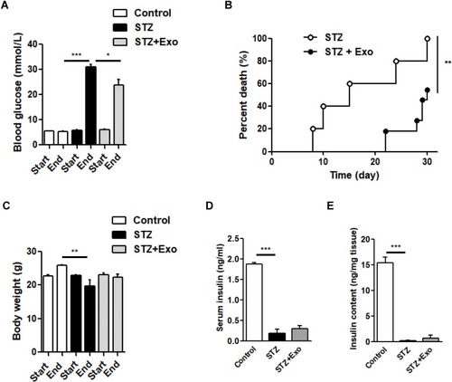
Figure 4 Effect of exosomes (Exo) on glucose metabolism in low-dose streptozotocin (STZ)-treated mice.
Notes: Male C57 mice were treated with 140mg/kg STZ, and Exo derived from MIN β-cells was transplanted at 10 days after STZ injection. (A-F) Intraperitoneal glucose tolerance tests (IPGTT) were performed on day 7 (A), day 17 (B) and day 35 (C). Area under curve (AUC) of IPGTT was calculated (D-F). Serum insulin levels before and after glucose load were detected at day 17 (G) and day 28 (H). (I) Insulin content extracted from pancreas were detected. Data were showed as mean ± SEM, *P<0.05, **P<0.01, ***P<0.001 between indicated groups, or between STZ + Exo and STZ-treated mice (Control, n=4; STZ, n=5; STZ+Exo, n=5).
Abbreviations: AUC, Area under curve; Exo, exosomes; IPGTT, intraperitoneal glucose tolerance test; STZ, streptozotocin.
Abbreviations: AUC, Area under curve; Exo, exosomes; IPGTT, intraperitoneal glucose tolerance test; STZ, streptozotocin.
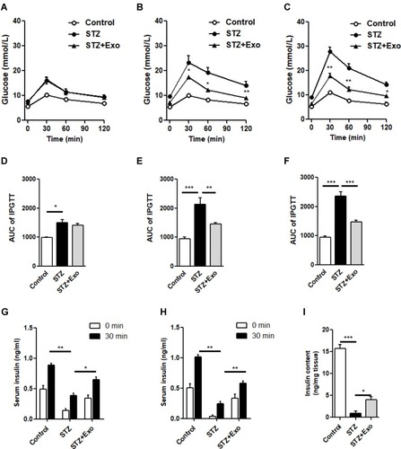
Figure 5 Effect of exosomes (Exo) on islet morphology of low-dose streptozotocin (STZ)-treated mice.
Notes: Mice were injected with 140mg/kg of STZ and transplanted with Exo after 10 days of STZ. The pancreatic sections from the mice were dual-stained for insulin in red and glucagon in green. DAPI staining blue indicated nuclei. Scale bar =20μm.
Abbreviations: Exo, exosomes; STZ, streptozotocin.
Abbreviations: Exo, exosomes; STZ, streptozotocin.
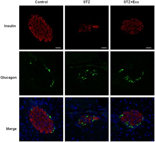
Figure 6 Effect of exosomes (Exo) on endothelial cell proliferation in islets.
Notes: Mice were injected with 140mg/kg of streptozotocin (STZ) and transplanted with Exo after 10 days of STZ. (A) The pancreas sections were dual-stained for insulin in red and CD31 in green. DAPI staining blue indicated nuclei. Scale bar =50 μm. Dashed line indicates the area of islets. (B) Quantification of CD31 density in islets, data were expression as ratio of CD31 density over islet area (12–16 islets/group). *P<0.05, ***P<0.001 between indicated groups.
Abbreviations: Exo, exosomes; STZ, streptozotocin.
Abbreviations: Exo, exosomes; STZ, streptozotocin.
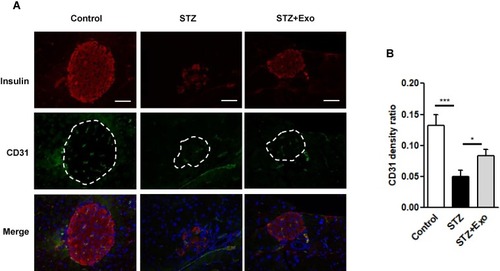
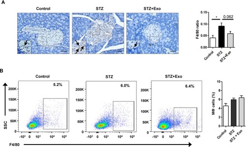
Figure 7 Effect of exosomes (Exo) on macrophage infiltration in mice treated with streptozotocin (STZ).
Notes: Mice were injected with 140mg/kg of STZ and transplanted with Exo after 10 days of STZ. (A) The pancreas sections were stained with F4/80, a marker for macrophages. Scale bar =50μm. Quantifications of F4/80-positive cells within islets were shown below. (B) Splenocytes were isolated from freshly obtained spleen, F4/80-positive cells were analyzed with flow cytometry. Data were showed as mean ± SEM, n=4. *P<0.05, between indicated groups.
Abbreviations: Exo, exosomes; STZ, streptozotocin.
Abbreviations: Exo, exosomes; STZ, streptozotocin.