Figures & data
Table 1 Primers Used in Quantitative Real-Time PCR
Figure 1 SMS protects ultrastructure integrity and attenuates cardiac fibrosis in diabetic rats. (A) Representative transmission electron micrographs of cardiac tissues (magnification = 15,000×). Scale bar (white) 50 μm; scale bar (black) 20 μm. (B) Representative images of myocardial tissue sections stained with Masson’s trichrome solution (magnification = 400×). Scale bar 50 μm. (C) Standard pictures of myocardial tissue sections stained with hematoxylin and eosin (magnification = 400×). Scale bar 50 μm.
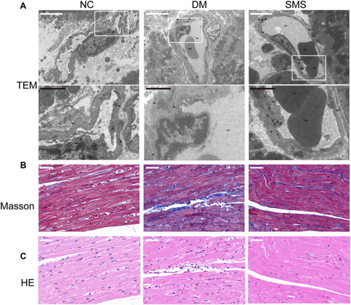
Figure 2 SMS protects cardiomyocytes from apoptosis induced by diabetes. (A) Standard pictures of myocardial tissue sections stained with TUNEL (magnification = 630×). Scale bar 100 μm. (B) Ratios of TUNEL-positive cells in different groups (n = 6 rats per group). Results are presented as means ± standard deviations. **p < 0.01 vs the NC group and ## p < 0.01 vs the DM group.
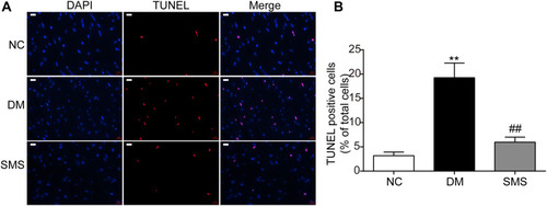
Figure 3 SMS prevents oxidative stress in the diabetic myocardium. Oxidative stress in the myocardial tissues was determined by measuring levels of (A) T-AOC, (B) 8-iso-PGF2α, and (C) 8-OHdG. Results are presented as means ± standard deviation. ***p < 0.001 vs the NC group and ### p < 0.001 vs the DM group (n = 6 rats per group).
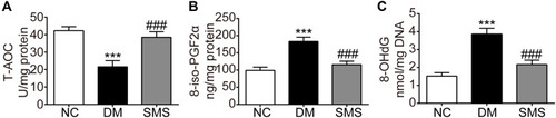
Figure 4 SMS blocks NOX activation in STZ-induced DM rats. (A and B) Real-time quantitative polymerase chain reaction and quantitative analysis of NOX2 and NOX4 mRNA levels in heart tissues. (C–F) Representative Western blot and quantitative analyses of NOX2 and NOX4 protein levels in heart tissues. β-actin was used as the internal control. Results are presented as means ± standard deviations. **p < 0.01 vs the NC group; # p < 0.05 vs the DM group, and ## p < 0.01 vs the DM group (n = 6 rats per group).
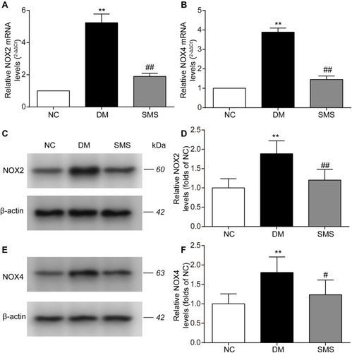
Figure 5 SMS inhibits NOX translocation in DM rats. The subcellular levels of p47phox and p67phox protein in heart tissues from different groups were detected by Western blot, and representative bands are shown in (A and D). The levels of cytomembrane and cytoplasm p47phox (B and C), and cytomembrane and cytoplasm p67phox (E and F) were normalized to the NC group. The results are presented as means ± standard deviations. **p<0.01 vs the NC group, # p<0.05 vs the DM group, and ## p<0.01 vs the DM group (n = 6 rats per group).
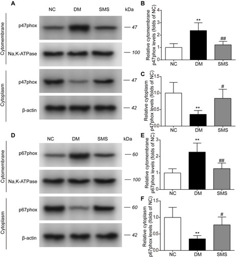
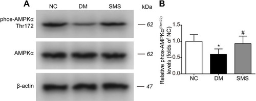
Figure 6 SMS restored AMPK activation in DM rats. The phosphorylation level of AMPKα in heart tissues from different groups was detected by Western blot assay. Representative bands were shown in (A) Phosphorylation levels of AMPKα (B) were normalized to NC. The results were presented as mean ± SD (n = 6). *p<0.05 vs the NC group, # p<0.05 vs the DM group.