Figures & data
Figure 1 Schematic representation of posterior retinal circulation. Parts of the blood supply to the optic disk, and the segmentation of the choroidal circulation are illustrated. The short posterior ciliary arteries (a–i) supply segments of the choroid (A–I) posterior to the equator of the globe.
©2006 Nature Publishing Group. Reproduced with permission from Aristodemou P, Stanford M. Therapy insight: the recognition and treatment of retinal manifestations of systemic vasculitis. Nat Clin Pract Rheumatol. 2006;2(8):443–451.Citation23
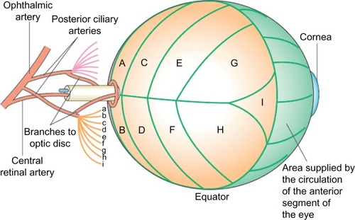
Table 1 Features of arteritic and nonarteritic anterior ischemic optic neuropathy
Figure 2 “Halo sign” of giant cell arteritis. Hypoechoic area (asterisk) around the temporal artery in longitudinal view (A) and transverse view (B) in a 70-year-old lady who presented with jaw pain and high inflammatory markers.
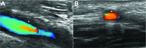
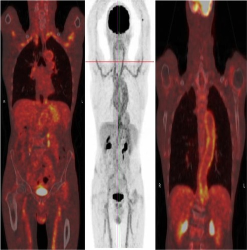
Figure 3 Fluorodeoxyglucose-positron emission tomographic scan showing florid uptake in large vessels including the aorta in a 74-year-old man who presented with polymyalgic symptoms and high inflammatory markers.