Figures & data
Figure 1 Nearly midline sagittal T1 MRI with gadolinium of pineal region germinoma in a 20-year-old male patient. The patient had signs and symptoms of double vision, light-near dissociation, and decreased upgaze. Cerebrospinal fluid had no markers of germinoma. Diagnosis was made with surgical resection via a infratentorial-supracerebellar approach to minimize risk of uncontrollable hemorrhage given the small size of bulky disease and proximity to major venous structures. (A) – splenium of corpus callosum; (B) – Vein of Galen; (C) – quadrigeminal cistern; (D) – inferior and superior colliculi; (E) – 4th ventricle; (F) – cerebral aqueduct (nearly collapsed); (G) – pineal germinoma, homogenously enhancing; (H) – subependymal spread of germinoma enhancing into mildly enlarged lateral ventricle; (I) – small sellar component of germinoma enhancing.
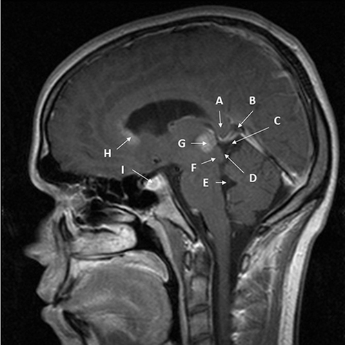
Figure 2 Midline sagittal T1 MRI with gadolinium of bifocal pineal region/suprasellar germinoma in a 15-year-old female patient. The patients had signs and symptoms of headache, bitemporal hemianopia, and polydipsia. Diagnosis was made via transsphenoidal resection of the suprasellar component of the mass to relieve the visual compromise. Arrows point out key structures as follows: (A) – splenium of corpus callosum; (B) – Vein of Galen; (C) – quadrigeminal cistern; (D) – inferior and superior colliculi; (E) – 4th ventricle; (F) – cerebral aqueduct; (G) – very small pineal germinoma, homogeneously enhancing; (H) - large sellar/suprasellar component of germinoma enhancing; (I) – 3rd ventricle; (J) – location of elevated and compressed optic chiasm (not well visualized).
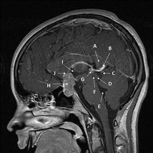
Figure 3 Axial CT without contrast of a 14-year-old boy presenting with headache and Parinaud’s syndrome. The tumor (A) is hyperdense, likely due to its cellularity. The third ventricle (B) is rounded, and the temporal horn of the lateral ventricle (C) is slightly dilated, consistent with early hydrocephalus.
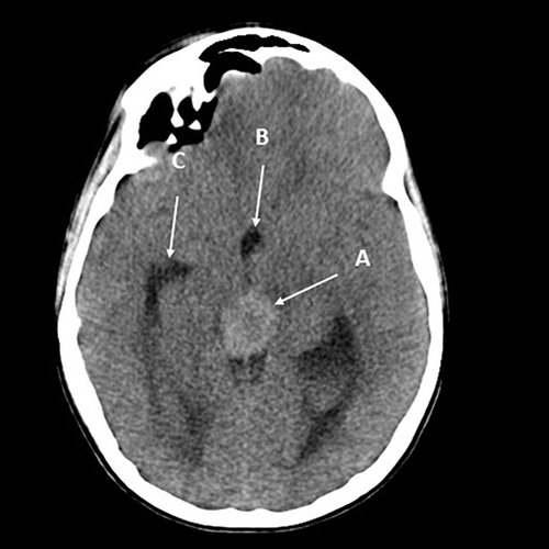
Table 1 Components in Diagnosis of Pineal Germinoma
Figure 4 Diagram of general management plan. Exceptions may occur. Option 1: four cycles of carboplatin on day 1 at 600 mg/m2 and etoposide at 150 mg/m2/day on days 1–3. Option 2: four cycles of carboplatin 600 mg/m2 on day 1 and VP16 90mg/m2 on days 1–3 alternating with ifosfamide 1800 mg/m2 on days 1–5 and VP16 90 mg/m2 on days 1–5.
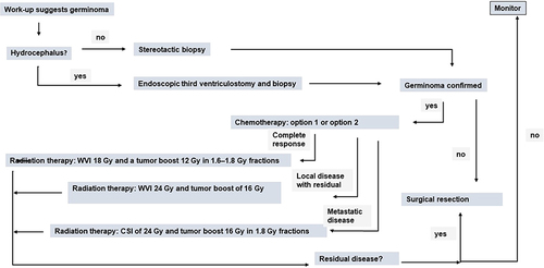
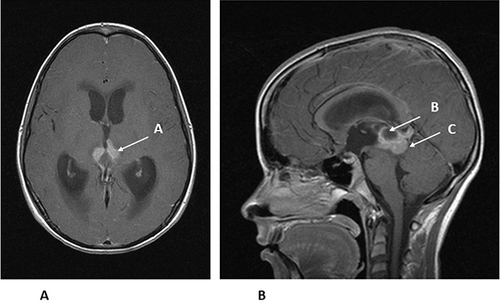
Figure 5 (A) Axial MRI with gadolinium of the same 14-year-old from . The enhancing tumor (A) is extending towards the thalamus. (B). Sagittal MRI with gadolinium of the same 14-year-old showing the enhancing tumor (B) and a cyst within the tumor (C). The patient had an endoscopic third ventriculostomy with biopsy to confirm the diagnosis of germinoma and treat his hydrocephalus. He was then treated with chemotherapy, followed by local radiation therapy.