856
Views
18
CrossRef citations to date
0
Altmetric
Review

Current Aspects in the Biology, Pathogeny, and Treatment of Candida krusei, a Neglected Fungal Pathogen

& ORCID Icon
Pages 1673-1689 | Published online: 10 Jun 2020

Figures & data

Figure 1 Candida krusei cell and colony morphology. (A) Yeast cells were grown in YPD broth until reach the exponential phase and then stained with calcofluor white, to label chitin. Scale bar = 10 µm. The arrowheads indicate the mother cells. (B) Cell filamentation was stimulated in RPMI medium incubated at 37°C. Scale bar = 20 µm. (C) A C. krusei colony grown on a YPD plate. Scale bar = 5.0 mm. Images from panels A and B were taken with a Zeiss Axioscope-40 microscope and an Axiocam MRc camera.

Figure 1 Candida krusei cell and colony morphology. (A) Yeast cells were grown in YPD broth until reach the exponential phase and then stained with calcofluor white, to label chitin. Scale bar = 10 µm. The arrowheads indicate the mother cells. (B) Cell filamentation was stimulated in RPMI medium incubated at 37°C. Scale bar = 20 µm. (C) A C. krusei colony grown on a YPD plate. Scale bar = 5.0 mm. Images from panels A and B were taken with a Zeiss Axioscope-40 microscope and an Axiocam MRc camera.

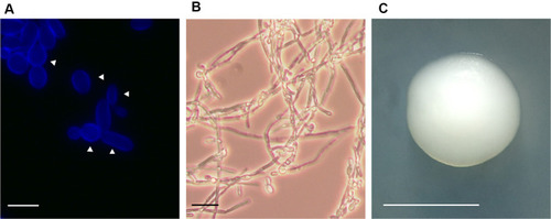
Table 1 Prediction of Some Virulence Factors in Candida krusei