Figures & data
Table 1 Primers Sets Used for nPCR Targeting COWP Gene
Table 2 Treatment Type and Dose Were Given to the Mice of T Subgroups
Table 3 Rate of Infection Throughout the Experiment
Table 4 Comparison of Means Oocysts/gm Stool Detected in All Groups at the End of the Experiment and Reduction Rate of Each Treatment
Table 5 Body Weights in All Groups Throughout the Experiment Compared to the Control Group
Table 6 IL10 Difference Among Groups Compared to Control
Table 7 TNF-α Difference Among Groups Compared to Control
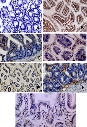
Figure 1 Immunohistochemical analysis of Cryptosporidium antigen in intestinal mice tissues of different groups: the control group and T1, T2, T3, T4, T5, and T6 were respectively represented in (A–G).
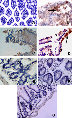
Figure 2 Immunohistochemical analysis of CD3 expression in intestinal mice tissues of different groups: the control group and T1, T2, T3, T4, T5, and T6 were represented in (A–G), respectively.