
Figures & data
Figure 1 Workflow of bacterial preparation from bacteria-positive blood cultures. After the liquid in the positive BC is mixed, a 6 mL aliquot is extracted with a syringe and transferred into a vacuum blood collection tube (with the red cap) containing coagulant (BD Vacutainer, USA). The centrifuge tube is gently mixed by inversion 5–6 times, and centrifuged at 350 × g for 15 min at room temperature. After centrifugation, the sediment is discarded. All the supernatant is transferred to a (yellow-capped) vacuum blood collection tube containing coagulant and separation gel (BD Vacutainer), and shaken gently to mix. It is then centrifuged for 5 min at 4500 × g at room temperature. After centrifugation, the supernatant is discarded and the sediment is carefully resuspended in 1 mL of saline solution with a micropipettor, and then all the liquid is transferred to a 1.5 mL microcentrifuge tube (Axygen, China). After centrifugation (150 × g, 5 min), the supernatant is transferred into a new 1.5 mL microcentrifuge tube and centrifuged for 5 min at 1500 × g. The supernatant is discarded and the pellet is resuspended in 1 mL of saline. The contents of the microcentrifuge tube are thoroughly mixed by vortexing for 5 s, and then centrifuged for 5 min at 1500 × g. The supernatant is discarded and the sediment is used for direct method tests.
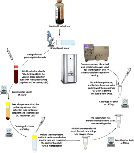
Table 1 Results of Direct Bacterial Identification with VITEK 2 Compact and MALDI-TOF MS Using the Developed Method
Table 2 Agreement and Errors in Antimicrobial Susceptibility Testing When Different Direct Methods Were Used
Table 3 Microorganism–Antimicrobial Combinations That Differed from Those Identified with the Standard Method
Table 4 Results of Extended-Spectrum β-Lactamase (ESBL) Testing with the Direct Method and the Standard Culture-Based Method
Table 5 Comparison of Direct and Standard Methods in Detecting Carbapenemases in Bacterial Strains