
Figures & data
Figure 1 (A) Diffuse low-density transparent areas can be seen in the lower part of the left femur and the upper part of the tibia. The boundary is unclear. The density of the patella is slightly uneven. The joint surface of the patella is narrow and rough, and the joint space is narrow. No abnormalities are found in the surrounding soft tissues. (B) Bone destruction is observed at up to a depth of approximately 2 cm inside the tibial tubercle, reaching deep into the medullary cavity.
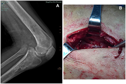
Figure 2 Microscopic morphology (400 x) of L-form NTM. (red spheroids, arrows).
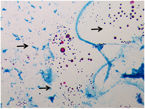
Figure 3 Morphology of L-form NTM (blue arrows) on electron microscopy.
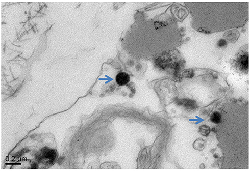
Figure 4 Acid-fast bacilli, slightly curved, with a tendency to branch (arrows) are observed on microscopy after the reversion test.
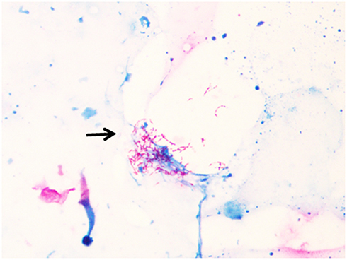
Table 1 Characteristics of Cases of NTM Osteomyelitis Identified in the Literature Review