Figures & data
Scheme 1 Illustration of BSA-Tpy NPs as a nanoplatform for photodynamic antibacterial therapy.
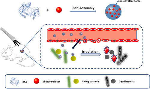
Figure 1 Characterizations of the BSA-Tpy. (A) Size distribution of NPs in the water. (B) TEM image. (C) The stability of BSA-Tpy. (D) Normalized FL intensity in water.
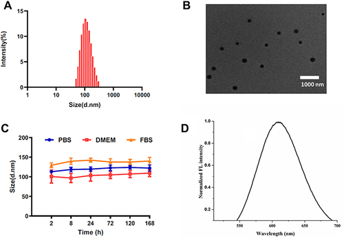
Figure 2 (A) The process and mechanism of ROS produced by materials under light irradiation. (B) Plotting of relative PL intensity (I/I0) at 525 nm versus the irradiation time.
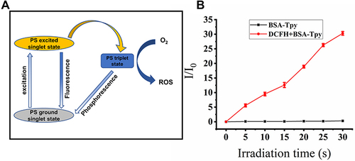
Figure 3 Cell viability of NIH/3T3 cell after being incubated with various concentrations of BSA, BSA-Tpy and Tpy for 24 h in the dark.
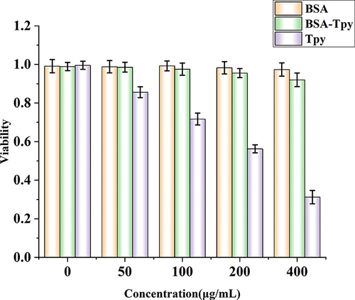
Figure 4 (A) Bright-field and fluorescent images of bacteria. E. coli, MRSA and were incubated with BSA-Tpy at pH 5.0 and for 25 min before imaging (Scale bar: 10 µm). (B) Image of plate counting method for E. coli and MRSA with of BSA-Tpy at pH 5.0.
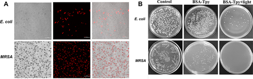
Figure 5 Visualizing BSA-Tpy NPs-induced morphological changes of bacteria, MRSA and E. coli for 25 min white light irradiation were imaged by SEM at pH 7.4 and pH 5.0 respectively, bacteria without treatment were set as controls.
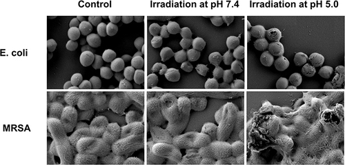
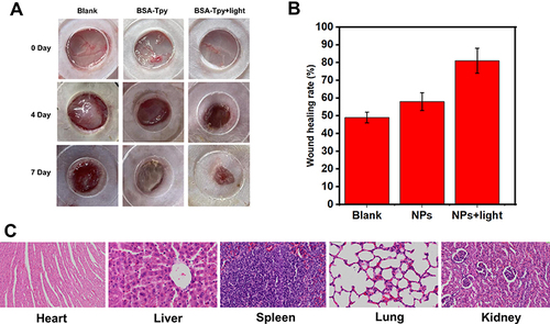
Figure 6 The evaluation of BSA-TpyNPs in treatment of bacteria-infected wounds of rats in vivo. (A) Images of MRSA-infected wounds after dealing with NPs with/without light ((50 mW·cm-2) on day 0 and day 4 postinfection. (B) The wounding healing rate of the MRSA-infected wound area on day 4 after the injury. (C) H&E staining of the organs in rats after treatment for 4 days.