1,176
Views
195
CrossRef citations to date
0
Altmetric
Review

Antimicrobial hydrogels: promising materials for medical application

, , , , , & show all
Pages 2217-2263 | Published online: 12 Apr 2018

Figures & data

Figure 1 The different applications of hydrogels.

Figure 1 The different applications of hydrogels.

Figure 2 Transmission electron microscope image of Escherichia coli cells treated with silver nanoparticles in liquid Luria-Bertani medium: (A) membrane of E. coli; (B) nanoparticles accumulated in the membrane and penetrated the cell (arrows).

Note: Reprinted from Adv Drug Deliv Rev. 65(13–14). Pelgrift RY, Friedman AJ, Nanotechnology as a therapeutic tool to combat microbial resistance1803–1815, Copyright (2013), with permission from Elsevier.Citation31

Figure 2 Transmission electron microscope image of Escherichia coli cells treated with silver nanoparticles in liquid Luria-Bertani medium: (A) membrane of E. coli; (B) nanoparticles accumulated in the membrane and penetrated the cell (arrows).Note: Reprinted from Adv Drug Deliv Rev. 65(13–14). Pelgrift RY, Friedman AJ, Nanotechnology as a therapeutic tool to combat microbial resistance1803–1815, Copyright (2013), with permission from Elsevier.Citation31

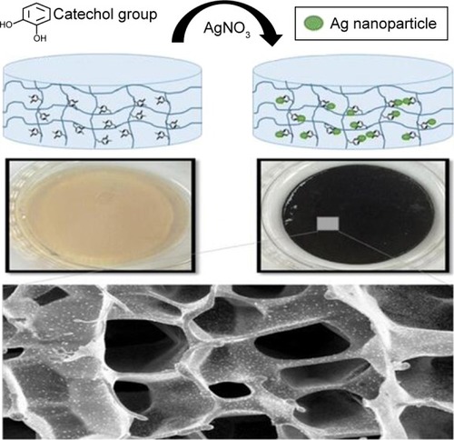
Table 1 Information of hydrogels with Ag NPs

Figure 3 Gao et al synthesized hydrogel containing Au NP-stabilized liposomes for antimicrobial application (A) illustrations of hydrogel containing nanoparticle-stabilized liposomes for topical antimicrobial delivery; (B) bacteria incubated with AuC–liposome hydrogel (PEGDMA 0.8 vol%) at pH = 4.5; (C) a zoomed-in image of (B).

Note: The scale bars in (B and C) represent 1 µm. Reproduced from Gao W, Vecchio D, Li J, et al. Hydrogel containing nanoparticle-stabilized liposomes for topical antimicrobial delivery. ACS Nano. 2014;8(3):2900–2907.Citation89

Figure 3 Gao et al synthesized hydrogel containing Au NP-stabilized liposomes for antimicrobial application (A) illustrations of hydrogel containing nanoparticle-stabilized liposomes for topical antimicrobial delivery; (B) bacteria incubated with AuC–liposome hydrogel (PEGDMA 0.8 vol%) at pH = 4.5; (C) a zoomed-in image of (B).Note: The scale bars in (B and C) represent 1 µm. Reproduced from Gao W, Vecchio D, Li J, et al. Hydrogel containing nanoparticle-stabilized liposomes for topical antimicrobial delivery. ACS Nano. 2014;8(3):2900–2907.Citation89

Table 2 Information of hydrogels with other metal nanoparticles

Figure 4 Multiple mechanisms of antimicrobial action of Ag NPs, ZnO NPs, copper-containing nanoparticles and Mg NPs are separately exhibited.

Note: Reprinted from Adv Colloid Interface Sci. 166(1–2). Dallas P, Sharma VK, Zboril R, Silver polymeric nanocomposites as advanced antimicrobial agents: classification, synthetic paths, appli cations, and perspectives, 119–135, Copyright 2011, with permission from Elsevier.37

Abbreviations: Ag NPs, silver nanoparticles; Mg NPs, magnesium-containing nanoparticles; NP, nanoparticle; ROS, reactive oxygen species; UV, ultraviolet; ZnO NPs, zinc oxide nanoparticles.

Figure 4 Multiple mechanisms of antimicrobial action of Ag NPs, ZnO NPs, copper-containing nanoparticles and Mg NPs are separately exhibited.Note: Reprinted from Adv Colloid Interface Sci. 166(1–2). Dallas P, Sharma VK, Zboril R, Silver polymeric nanocomposites as advanced antimicrobial agents: classification, synthetic paths, appli cations, and perspectives, 119–135, Copyright 2011, with permission from Elsevier.37Abbreviations: Ag NPs, silver nanoparticles; Mg NPs, magnesium-containing nanoparticles; NP, nanoparticle; ROS, reactive oxygen species; UV, ultraviolet; ZnO NPs, zinc oxide nanoparticles.

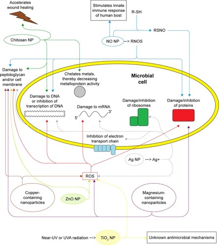
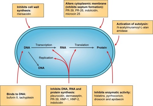
Table 3 Antimicrobial mechanism of nanoparticles

Figure 5 Development of antibiotics and appearance of drug resistance are summarized chronologically referring to Huh and Kwon,Citation35 Andersson and Hughes,Citation132 Rodriguez- Rojas et al,Citation133 van Hoek et al,Citation134 Molton et al.135

Abbreviations: E. coli, Escherichia coli; K. pneumoniae, Klebsiella pneumoniae; MRSA, methicillin resistant S. aureus; S. aureus, Staphylococcus aureus; VISA, vancomycin intermedicate resistant S. auereus; VRE, vancomycin-resistant Enterococcus; VRSA, vancomycin-resistant S. aureus.

Figure 5 Development of antibiotics and appearance of drug resistance are summarized chronologically referring to Huh and Kwon,Citation35 Andersson and Hughes,Citation132 Rodriguez- Rojas et al,Citation133 van Hoek et al,Citation134 Molton et al.135Abbreviations: E. coli, Escherichia coli; K. pneumoniae, Klebsiella pneumoniae; MRSA, methicillin resistant S. aureus; S. aureus, Staphylococcus aureus; VISA, vancomycin intermedicate resistant S. auereus; VRE, vancomycin-resistant Enterococcus; VRSA, vancomycin-resistant S. aureus.

Table 4 Information of hydrogels with antibiotic agents

Figure 6 Mode of action for intracellular antimicrobial peptide activity. In this figure Escherichia coli was shown as the target microorganism from Brogden.

Note: Reprinted by permission from Springer Nature, Nat Rev Microbiol, Brogden KA, Antimicrobial peptides: pore formers or metabolic inhibitors in bacteria? 2005;3(3): 238–250, Copyright 2005.Citation235

Figure 6 Mode of action for intracellular antimicrobial peptide activity. In this figure Escherichia coli was shown as the target microorganism from Brogden.Note: Reprinted by permission from Springer Nature, Nat Rev Microbiol, Brogden KA, Antimicrobial peptides: pore formers or metabolic inhibitors in bacteria? 2005;3(3): 238–250, Copyright 2005.Citation235

Figure 7 Morphological observation of various microorganisms seeded on hydrogels by scanning electron microscope. Left columns (control), right columns (antimicrobial hydrogels).

Note: Reprinted from Biomaterials. 32(11). Zhou C, Li P, Qi X, et al, A photopolymerized antimicrobial hydrogel coating derived from epsilon-poly-l-lysine, 2704–2712, Copyright 2011, with permission from Elsevier.242

Abbreviations: C. albicans, Candida albicans; E. coli, Escherichia coli; F. solani, Fusarium solani; P. aeruginosa, Pseudomonas aeruginosa; S.aureus, Staphylococcus aureus; S. marcescens, Serratia marcescens.

Figure 7 Morphological observation of various microorganisms seeded on hydrogels by scanning electron microscope. Left columns (control), right columns (antimicrobial hydrogels).Note: Reprinted from Biomaterials. 32(11). Zhou C, Li P, Qi X, et al, A photopolymerized antimicrobial hydrogel coating derived from epsilon-poly-l-lysine, 2704–2712, Copyright 2011, with permission from Elsevier.242Abbreviations: C. albicans, Candida albicans; E. coli, Escherichia coli; F. solani, Fusarium solani; P. aeruginosa, Pseudomonas aeruginosa; S.aureus, Staphylococcus aureus; S. marcescens, Serratia marcescens.

Table 5 Information of hydrogels with inherent antibacterial activity

Figure 8 A new strategy that uses catecholic chemistry to synthesize antimicrobial silver nanoparticles impregnated into antifouling zwitterionic hydrogels.

Notes: On the top is the schematic illustration of the combination of AgNPs and antifouling hydrogel. In the middle, Photographs show the changes in color of hydrogels by changing the pH because of reaction that converts the Ag+ into solid AgNPs. The bottom section shows the surface structure and the morphology of hydrogel via scanning electron microscopy. Reprinted with permission from GhavamiNejad A, Park CH, Kim CS. In situ synthesis of antimicrobial silver nanoparticles within antifouling zwitterionic hydrogels by catecholic redox chemistry for wound healing application. Biomacromolecules. 2016;17(3):1213–1223. Copyright (2016), American Chemical Society.Citation287

Figure 8 A new strategy that uses catecholic chemistry to synthesize antimicrobial silver nanoparticles impregnated into antifouling zwitterionic hydrogels.Notes: On the top is the schematic illustration of the combination of AgNPs and antifouling hydrogel. In the middle, Photographs show the changes in color of hydrogels by changing the pH because of reaction that converts the Ag+ into solid AgNPs. The bottom section shows the surface structure and the morphology of hydrogel via scanning electron microscopy. Reprinted with permission from GhavamiNejad A, Park CH, Kim CS. In situ synthesis of antimicrobial silver nanoparticles within antifouling zwitterionic hydrogels by catecholic redox chemistry for wound healing application. Biomacromolecules. 2016;17(3):1213–1223. Copyright (2016), American Chemical Society.Citation287

Figure 9 Graphical representation of MICs obtained after growing S. aureus and P. aeruginosa in the presence of different concentrations of gentamicin and ZnO/gentamicin–chitosan.

Note: Reprinted from Int J Pharm. 463(2). Vasile BS, Oprea O, Voicu G, et al, Synthesis and characterization of a novel controlled release zinc oxide/gentamicin-chitosan composite with potential applications in wounds care, 161–169, Copyright 2014, with permission from Elsevier.Citation290

Abbreviations: MICs, minimal inhibition concentrations; S. aureus, Staphylococcus aureus; P. aeruginosa, Pseudomonas aeruginosa.

Figure 9 Graphical representation of MICs obtained after growing S. aureus and P. aeruginosa in the presence of different concentrations of gentamicin and ZnO/gentamicin–chitosan.Note: Reprinted from Int J Pharm. 463(2). Vasile BS, Oprea O, Voicu G, et al, Synthesis and characterization of a novel controlled release zinc oxide/gentamicin-chitosan composite with potential applications in wounds care, 161–169, Copyright 2014, with permission from Elsevier.Citation290Abbreviations: MICs, minimal inhibition concentrations; S. aureus, Staphylococcus aureus; P. aeruginosa, Pseudomonas aeruginosa.