Figures & data
Figure 1 A schematic illustration of nanostructured Lipid Carrier (NLC) on right and solid lipid nanoparticles (SLN) on left
Notes: Reproduced from Hsu CY, Wang PW, Alalaiwe A, Lin ZC, Fang JY. Use of lipid Nanocarriers to improve Oral delivery of vitamins. Nutrients. 2019;11(1):68-97Citation325
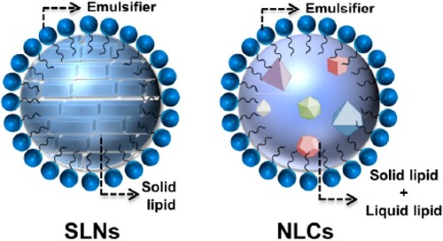
Table 1 Nanocarrier Encapsulated Herbal Formulations
Figure 2 A schematic illustration of oil (O) in water (W) nanoemulsion.
Notes: Reproduced from Agnihotri N, Soni GC, Chanchal DK, Tiwari S. A Scientific Review On Nanoemulsion For Targeting Drug Delivery System. Int J Life Sci Rev. 2019;5(2):16-29Citation326
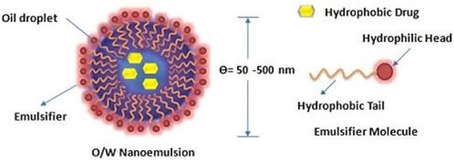
Figure 3 A schematic illustration of silver-loaded titanium dioxide nanocapsule.
Notes: Adapted from Hérault N, Wagner J, Abram SL, et al. Silver-Containing Titanium Dioxide Nanocapsules for Combating Multidrug-Resistant Bacteria. Int J Nanomed. 2020;15:1267-1281Citation327
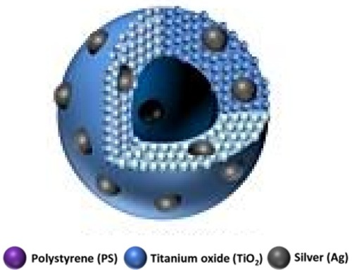
Figure 4 A schematic illustration of Polyethylene glycate (PEG)-aptamer-liposome-doxorubicin (DOX); a type of lipid drug-conjugate.
Notes: Reproduced from Dou XQ, Wang H, Zhang J, et al. Aptamer–drug conjugate: targeted delivery of doxorubicin in a HER3 aptamer-functionalized liposomal delivery system reduces cardiotoxicity. Int J Nanomed. 2018;13:763-776Citation328
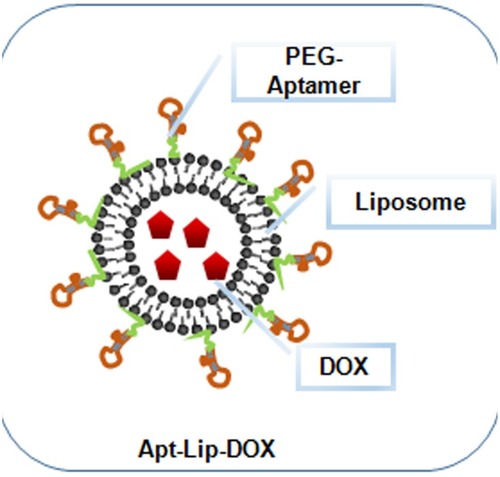
Figure 5 A schematic illustration of liposome (A), transferosome (B), niosome (C) and ethosome (D).
Notes: Adapted with permission from Frontier in Pharmacology. Sercombe L, Veerati T, Moheimani F, Wu SY, Sood AK, Hua S. Advances and challenges of liposome assisted drug delivery. Front. Pharmacol. 2015;6:286.Citation324
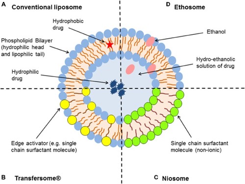
Figure 6 A schematic illustration of dendrimer.
Notes: Reproduced from ud Din F, Aman W, Ullah I, et al. Effective use of nanocarriers as drug delivery systems for the treatment of selected tumors. Int J Nanomed. 2017;12:7291-7309Citation8
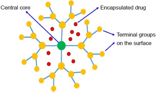
Figure 7 A schematic illustration of micelle.
Notes: Reproduced from ud Din F, Aman W, Ullah I, et al. Effective use of nanocarriers as drug delivery systems for the treatment of selected tumors. Int J Nanomed. 2017;12:7291-7309Citation8
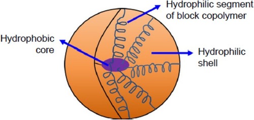
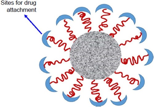
Figure 8 A schematic illustration of nanosphere.
Notes: Reproduced from Harper 3D.Citation142
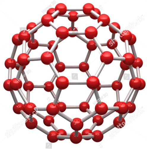
Figure 9 A schematic illustration of fentofibrate nanocrystals (FNB-NCs).
Notes: Reproduced from Kevadiya BD, Chen L, Zhang L, Thomas MB, Davé RN. Fenofibrate Nanocrystal Composite Microparticles for Intestine-Specific Oral Drug Delivery System. Pharmaceuticals. 2019;12(3):109-124Citation329
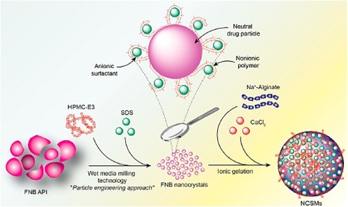
Figure 10 A schematic illustration of phytosome.
Notes: Reproduced Karthivashan G, Masarudin MJ, Kura AU, Abas F, Fakurazi S. Optimization, formulation, and characterization of multiflavonoids-loaded flavanosome by bulk or sequential technique. Int J Nanomed. 2016;11:3417-3434Citation169
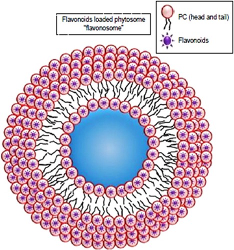
Figure 11 A schematic illustration of APC-SNEDDS dissolved in distilled water. APC: Akebia saponin D-phospholipid complex.
Notes: Reproduced from Shen J, Bi J, Tian H, et al. Preparation and evaluation of a self-nanoemulsifying drug delivery system loaded with akebia saponin D–phospholipid complex. Int J Nanomed. 2016;11:4919-4929Citation183
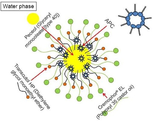
Figure 12 A schematic illustration of SMEDDS.
Notes: Adapted from Quan G, Niu B, Singh V, et al. Supersaturable solid self-microemulsifying drug delivery system: precipitation inhibition and bioavailability enhancement. Int J Nanomed. 2017;12:8801-8811Citation330
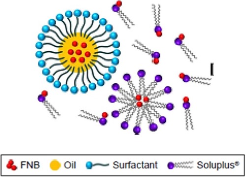
Figure 13 A schematic illustration of dexamethasone loaded nanofibers (Dex-NS).
Notes: Adapted from Lee JW, Lee HY, Park SH, et al. Preparation and evaluation of dexamethasone-loaded electrospun nanofiber sheets as a sustained drug delivery system. Materials. 2016;9(3):175-186Citation331
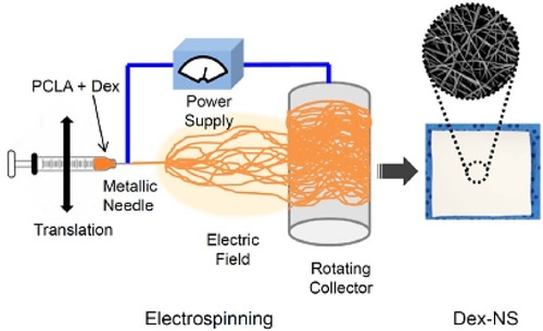
Figure 14 A schematic illustration of polymerosome.
Notes: Adapted from Prabhu RH, Patravale VB, Joshi MD. Polymeric nanoparticles for targeted treatment in oncology: current insights. Int J Nanomed. 2015;10:1001-1018Citation84
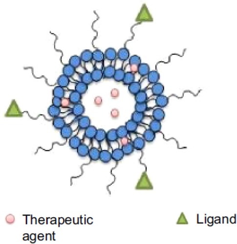
Figure 15 Transmission electron micrographs of 20(S)-protopanaxadiol cubosome with (A) and without (B) Pierine.
Notes: Reproduced from Jin X, Zhang ZH, Sun E, et al. Enhanced oral absorption of 20 (S)-protopanaxadiol by self-assembled liquid crystalline nanoparticles containing piperine: in vitro and in vivo studies. Int J Nanomed. 2013;8:641-652Citation332
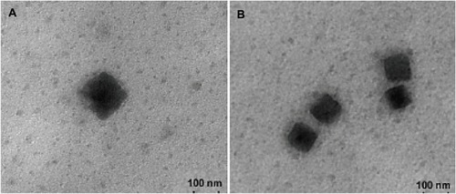
Figure 16 A schematic illustration of chitosan nanoparticle.
Notes: Reproduced from Tan Q, Liu W, Guo C, Zhai G. Preparation and evaluation of quercetin-loaded lecithin-chitosan nanoparticles for topical delivery. Int J Nanomed. 2011;6:16211630.Citation233
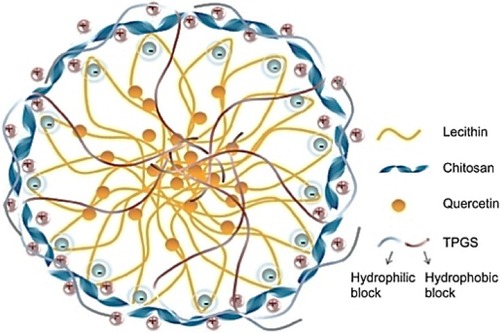
Figure 17 A schematic illustration of biopolymeric hydrogel.
Notes: Reproduced with permission from MDPI. Zhao F, Yao D, Guo R, Deng L, Dong A, Zhang J. Composites of 2075 polymer hydrogels and nanoparticulate systems for biomedical and pharmaceutical applications. Nanomaterials. 2015;5(4):2054–2130.Citation242
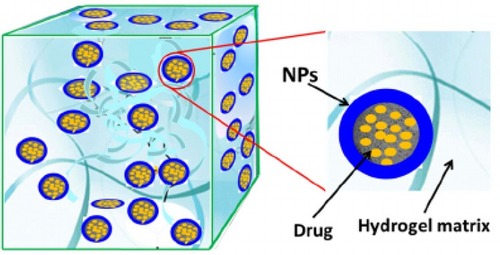
Figure 18 A schematic illustration of biopolymeric drug conjugate.
Notes: Reproduced from Safer AM, Leporatti S, Jose J, Soliman MS. Conjugation Of EGCG And Chitosan NPs As A Novel Nano-Drug Delivery System. Int J Nanomed. 2019;14:8033-8046.Citation333
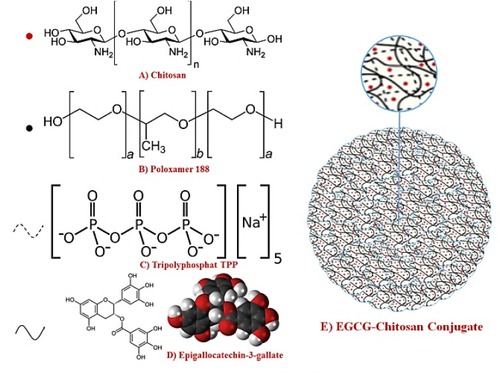
Figure 19 A schematic illustration of gold nanoparticle.
Notes: Reproduced with permission from Luna Nanotech.Citation260
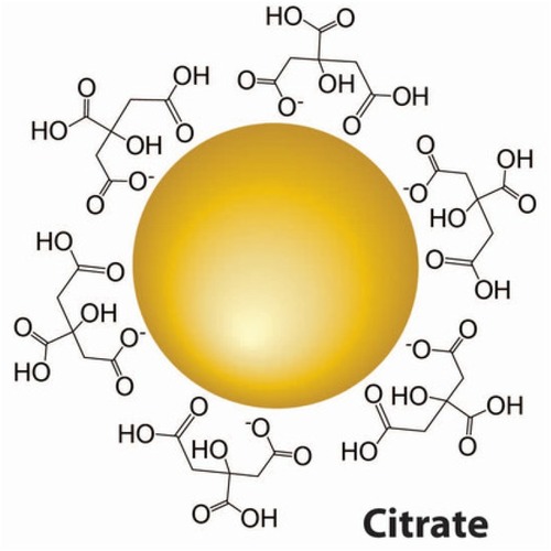
Figure 20 A schematic illustration of silica nanoparticle.
Notes: Reproduced from ud Din F, Aman W, Ullah I, et al. Effective use of nanocarriers as drug delivery systems for the treatment of selected tumors. Int J Nanomed. 2017;12:7291-7309.Citation8
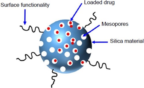
Figure 21 A schematic illustration of magnetic nanoparticle.
Notes: Adapted with permission from Frontier in Microbiology. Souza AC, Amaral AC. Antifungal therapy for systemic mycosis and the nanobiotechnology era: improving efficacy, biodistribution and toxicity. Front Microbiol. 2017;8(336):1–13. Citation280
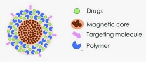
Figure 22 A schematic illustration of veteran cockle shell-derived calcium carbonate nanoparticles.
Notes: Reproduced from Muhammad Mailafiya M, Abubakar K, Danmaigoro A, et al. Cockle Shell-Derived Calcium Carbonate (Aragonite) Nanoparticles: A Dynamite to Nanomedicine. Appl Sci. 2019 ;9(14):2897-2922.Citation334
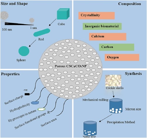
Figure 23 A schematic illustration of halloysite clay nanotubes.
Notes: Reproduced with permission from Kamal N, Kochkodan V, Zekri A, Ahzi S. Polysulfone Membranes Embedded with Halloysites Nanotubes: Preparation and Properties. Membranes. 2020;10(1):2-29.Citation335
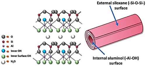
Figure 24 A schematic illustration of single walled carbon nanotube (A) and double walled carbon nanotube (B).
Notes: Reproduced from ud Din F, Aman W, Ullah I, et al. Effective use of nanocarriers as drug delivery systems for the treatment of selected tumors. Int J Nanomed. 2017;12:7291-7309.Citation8
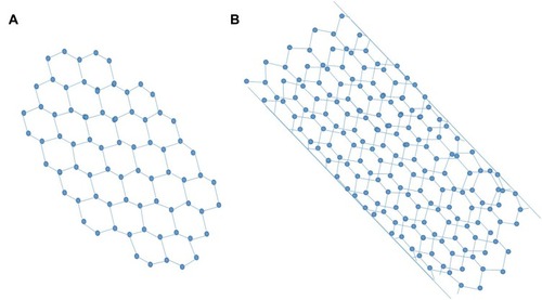
Figure 25 Types and structures of hybrid nanocarrier.
Notes: Adapted from Prabhu RH, Patravale VB, Joshi MD. Polymeric nanoparticles for targeted treatment in oncology: current insights. Int J Nanomed. 2015;10:1001-1018.Citation84
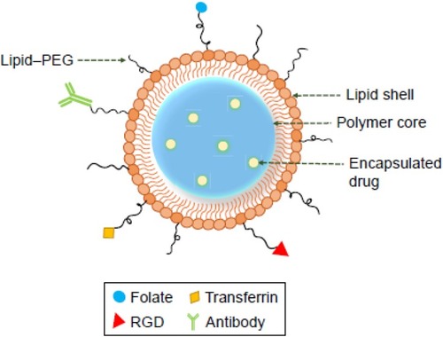
Figure 26 A schematic illustration of biological nanocarrier.
Notes: Reproduced from ud Din F, Aman W, Ullah I, et al. Effective use of nanocarriers as drug delivery systems for the treatment of selected tumors. Int J Nanomed. 2017;12:7291-7309.Citation8