Figures & data
Table 1 Advantages and Limitations of Some Remarkable Published Studies About Functionalized Graphene Oxide Nanoparticles
Figure 1 Schematic of (A) cRGD conjugated to chitosan (RC) and (B) RC-GO-Dox nanocarrier.
Note: Copyright ©2014. Elsevier. Reproduced from Wang C, Chen B, Zou M, Cheng G. Cyclic RGD-modified chitosan/graphene oxide polymers for drug delivery and cellular imaging. Colloids and Surfaces B: Biointerfaces. 2014; 122: 332–340.Citation61
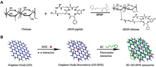
Figure 2 Scheme of TRAIL/DOX-fGO nanoplatform. (A) Main components of TRAIL/DOX-fGO, and (B) Step by step activity of TRAIL/DOX-fGO, from vessel administration to drug release in the cell nucleus.
Note: Copyright ©2015. John Wiley and Sons. Reproduced from Jiang T, Sun W, Zhu Q, et al. Furin‐Mediated Sequential Delivery of Anticancer Cytokine and Small‐Molecule Drug Shuttled by Graphene. Advanced Materials. 2015; 27 (6):1021–1028.Citation63
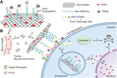
Figure 3 (A) NGO-SS-mPEG in extracellular environment (low GSH level), (B) it is internalized into the tumor cell (high GSH level), (C) linkage is cleaved, and (D) DOX is released.
Note: Copyright ©2012. John Wiley and Sons. Reproduced from Wen H, Dong C, Dong H, et al. Engineered redox‐responsive PEG detachment mechanism in PEGylated nano‐graphene oxide for intracellular drug delivery. Small. 2012; 8 (5): 760–769.Citation81
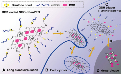
Figure 4 The mechanism of DOX release process in acidic and high GSH level conditions from rGO/QC-PEG and rGO/QC-PEG/Plu-SH.
Note: Copyright ©2013. Elsevier. Reproduced from Al-Nahain A, Lee SY, In I, et al. Triggered pH/redox responsive release of doxorubicin from prepared highly stable graphene with thiol grafted pluronic. Int J Pharm. 2013; 450 (1): 208–217.Citation84
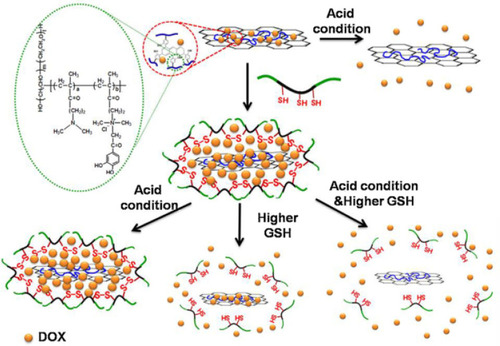
Figure 5 (A) rGO-AuNRVe morphology by TEM, (B) Dox release process from rGO-AuNRVe in the cell (Reproduced with permission).
Note: Copyright ©2015. American Chemical Society. Reproduced from Song J, Yang X, Jacobson O, et al. Sequential drug release and enhanced photothermal and photoacoustic effect of hybrid reduced graphene oxide-loaded ultrasmall gold nanorod vesicles for cancer therapy. ACS nano. 2015; 9 (9): 9199–9209.Citation119
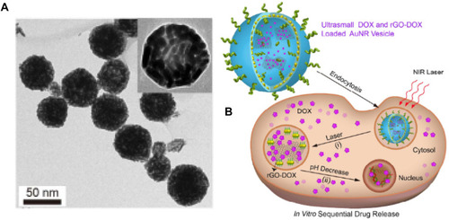
Figure 6 (A) Preparation of Dox loaded GOF-Lip-FA nano theranostic system in a schematic view, (B) Targeted cellular uptake of the GOF-Lip-FA nanohybrid and DOX release in cells body (red particles), (C) In vivo biodistribution of the platform by IV injection and cancer cells uptake, and (D) near-infrared 4T1 breast tumor cells death.
Note: Copyright ©2019. American Chemical Society. Reproduced from Prasad R, Yadav AS, Gorain M, et al. Graphene Oxide Supported Liposomes as Red Emissive Theranostics for Phototriggered Tissue Visualization and Tumor Regression. ACS Applied Bio Materials. 2019; 2 (8): 3312–3320.Citation168
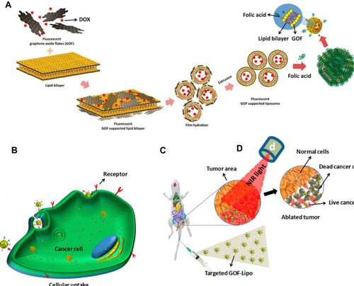
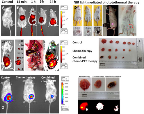
Figure 7 In vivo performance of GOF-Lip-FA nano theranostic platform. (A–C) in vivo 4T1 cancer diagnosis and distribution of nano theranostic systems by near-infrared fluorescence imaging. (D–E) images of the tumor-bearing animal under laser irradiation and tumor size shrinkage shown in dotted circles. (F) Tumor size after different therapeutic paths. (G) The decrease in tumor size after 21 days. (H) near-infrared fluorescence images of tumor regression after various treatments.
Note: Copyright ©2019. American Chemical Society. Reproduced from Prasad R, Yadav AS, Gorain M, et al. Graphene Oxide Supported Liposomes as Red Emissive Theranostics for Phototriggered Tissue Visualization and Tumor Regression. ACS Applied Bio Materials. 2019; 2 (8): 3312–3320.Citation168