Figures & data
Figure 1 Schematic representation of the aptamer-based capture platform to identify the pathogen in human blood.
Notes: Adapted from Shen H, Wang J, Liu H, et al. Rapid and Selective Detection of Pathogenic Bacteria in Bloodstream Infections with Aptamer-Based Recognition, ACS Appl. Mater. Interfaces. 2016; 8 (30): 19371–19378. Copyright © 2016, with permission from American Chemical Society.Citation30
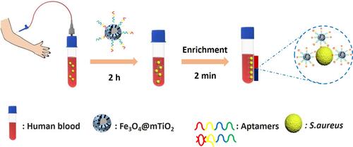
Figure 2 Design of organic-inorganic nanoflowers composites for integration in electrochemical detection of E. coli.
Notes: Reprinted from Li Y, Xe G, Qu J, et al. A new biosensor based on the recognition of phages and the signal amplification of organic-inorganic hybrid nanoflowers for discriminating and quantitating live pathogenic bacteria in urine. Sensors and Actuators B: Chemical 2018; 258 (30): 803–812. Copyright © 2017, with permission from Elsevier.Citation40
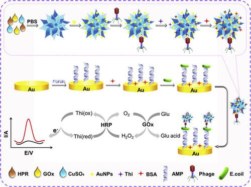
Table 1 Designs of MSNs Employed in MSN Integrated Biosensors for Detection of Bacterial Infections
Figure 3 (A) Co-condensation of luminophores via covalent bonding and noncovalent interaction in mesoporous siliceous frameworks. (B) Post-synthesis approaches for the addition of optical units into MSN.
Notes: (A) Reprinted from Gao M, Zeng J, Liang K, Zhao D, Kong B. Interfacial Assembly of Mesoporous Silica‐Based Optical Heterostructures for Sensing Applications. Adv. Funct. Mater. 2020, 1906950. Copyright 2020 with permission from WILEY‐VCH.Citation81
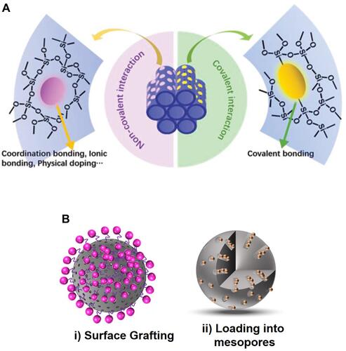
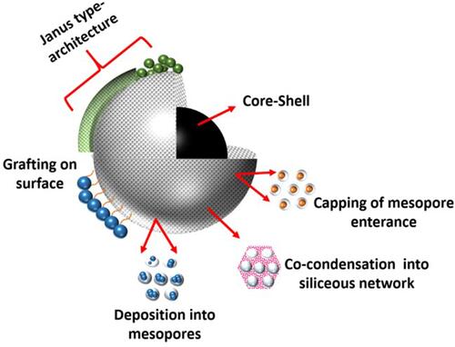
Figure 4 Strategies for incorporation of inorganic species into MSN.