Figures & data
Figure 1 Schematic illustration of the classification (A), surface modification and functionalization (B and C), synthesis and formation mechanism (D), and application of mesoporous silica nanomaterials (E and F) in this review.
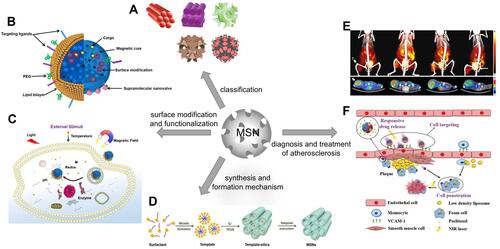
Table 1 Classification and Characteristics of Common Mesoporous Silica Nanomaterials
Figure 2 A schematic diagram of the synthesis of MSNs.
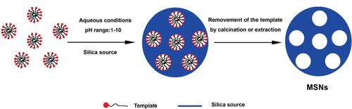
Figure 3 Schematic diagrams of the formation mechanism of mesoporous silica. (A) Scheme of the formation mechanism of MCM-41. (B) SANS method. (C) “expansion and contraction” mechanism.
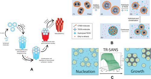
Figure 4 A schematic diagram of surface modification of mesoporous silica. (A) amino functionalization. (B) polymer modification.
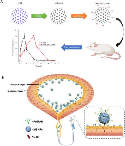
Table 2 Summary of Application of Mesoporous Silica in Atherosclerosis
Figure 5 Applications of mesoporous silica in the diagnosis and treatment of AS. (A) A schematic diagram of an effective macrophage tracking protocol based on an in vivo bioorthogonal F-18 labeling reaction using the PET system. (B) Schematic illustration of identifying macrophage enrichment in atherosclerotic plaques by dual-modal US imaging and MRI based on biodegradable CD68-Fe-HSN. (C) Schematic illustration of the synthesis process of MJAMS/PTX/aV and the mechanism of treatment of AS using the MJAMS/PTX/aV coated balloon.
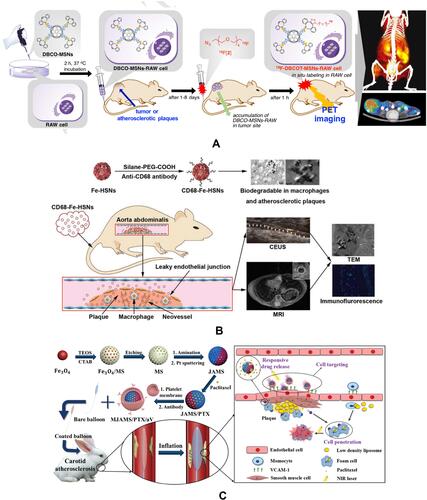
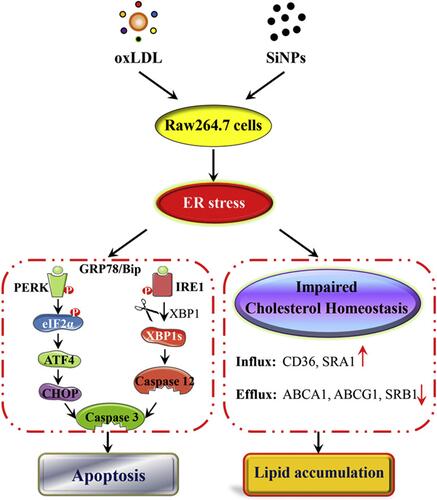
Figure 6 The induction of foam cell along with the disturbance on cholesterol influx/efflux balance, and promotion of apoptosis via ER stress PERK/eIF2α/ATF4 and IRE1α/XBP1 signaling cascade by SiNPs and oxLDL coexposure in a macrophage model.