Figures & data

Figure 1 Schematic demonstration and possible synthesis mechanism for the green synthesized VB-Au NPs with the leaves extract of Viola betonicifolia.
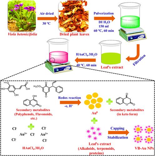
Figure 2 UV-Visible spectra of (A) leave extract of Viola betonicifolia and (B) green synthesized VB-Au NPs. FTIR spectra of (C) leave extract of Viola betonicifolia and (D) green synthesized VB-Au NPs.
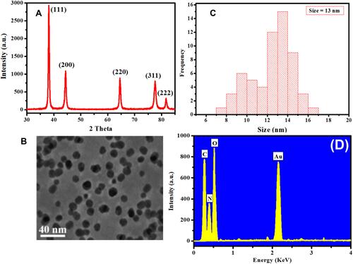
Figure 3 (A) XRD pattern, (B) TEM image, (C) size distribution, and (D) EDX pattern for the green synthesized VB-Au NPs.
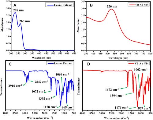
Figure 4 Antibacterial activity of VB-Au NPs in terms of (A) log10 reduction (p < 0.0004) and (B) % killing efficiency of bacterial strains in comparison to leaves extract of Viola betonicifolia and CH-Au NPs (p < 0.0004). (C) Representative images of control and treated E. coli and P. aeruginosa with VB-Au NPs.
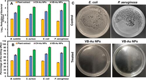
Figure 5 CLSM images of B. subtilis (A), S. aureus (B), E. coli (C), and P. aeruginosa (D). Green and red represent alive and dead bacterial cells, respectively.
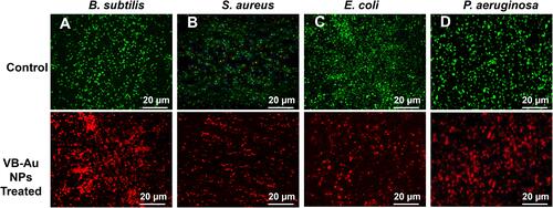
Figure 6 CLSM images of ROS generation in untreated (control) and treated bacteria with hydrogen peroxide (H2O2) and synthesized VB-Au NPs.
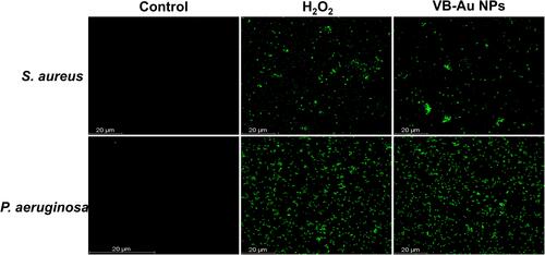
Figure 7 Comparison of Gram-negative and Gram-positive bacterial cell wall and proposed antibacterial mechanism of green synthesized VB-Au NPs. Created with BioRender.com.
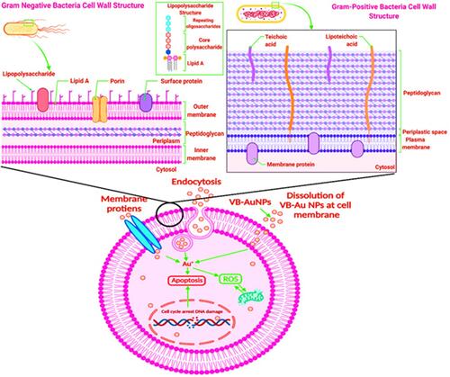
Figure 8 Antifungal activity of VB-Au NPs in terms of (A) log10 reduction (p < 0.0008) and (B) % killing efficiency (p < 0.0008) of fungal strains in comparison to leaves extract of Viola betonicifolia and CH-Au NPs.
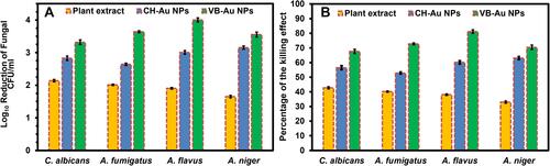
Figure 9 Biofilm inhibition performance of the synthesized VB-Au NPs against the (A) bacterial (p < 0.006) and (B) fungal strains (p < 0.004) in comparison to Viola betonicifolia leaves extract and CH-Au NPs.
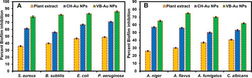
Figure 10 Cytotoxic potential in terms of cell viability percentage against MCF-7 carcinoma cells treated with Viola betonicifolia leaves extract, CH-Au NPs, and VB-Au NPs (p < 0.0001).
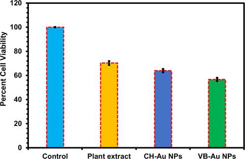
Figure 11 CLSM images of MCF-7 cells before (A) control and after incubating with (B) plant extract, (C) CH-Au NPs and (D) VB-Au NPs.
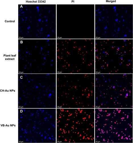
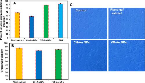
Figure 12 (A) Antioxidant activity of the newly synthesized VB-Au NPs in comparison to Viola betonicifolia leaves extract, CH-Au NPs, and external standard (BHT) (p < 0.0001). (B) Biocompatibility analysis of the newly synthesized VB-Au NPs with hMSCs compared to the leaves extract of Viola betonicifolia and CH-Au NPs (p < 0.0002). (C) Inverted microscopic images of the hMSCs treated with plant extract, CH-Au NPs, and VB-Au NPs.