Figures & data
Figure 1 Preparation of GA/WS-CQDs hydrogels. (A) The schematic diagram of the GA/WS-CQDs-gel preparation. (B) FT-IR characterization. (C) SEM images of the GA-gel and GA/WS-CQDs-gel. (D–G) Rheology test of the GA-gel and GA/WS-CQDs-gel. Frequency scan (D), Amplitude scan (E), Cycle scan (F), Complex viscosity scan (G). (H) Schematic of GA/WS-CQDs-gel injectability. (I) Self-healing process of GA/WS-CQDs-gel after cutting.
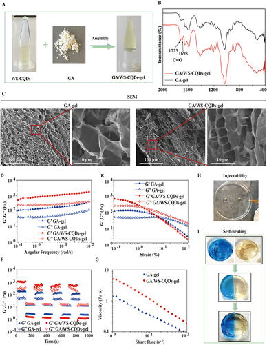
Table 1 The Synergistic Interaction of Combined WS-CQDs with GA Against MRSA
Figure 2 The noticeable anti-MRSA synergism between WS-CQDs and GA. (A and B) Heat map visually displayed the growth inhibition of bacterial treated with different combination between WS-CQDs and GA. The numbers in the heat map are the corresponding OD values. (C and D) Standard plate counting method, the representative digital images of agar plates displaying results for WS-CQDs, GA, GA/WS-CQDs and PBS (control group) against MRSA. Combination 1 (low concentration of GA and high concentration of WS-CQDs), combinations 2 (1/2 × MICs of GA and 1/2 × MICs of WS-CQDs), and combinations 3 (high concentration of GA and low concentration of WS-CQDs).
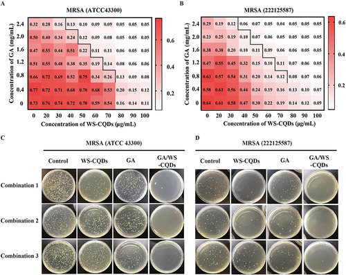
Figure 3 In vitro the cumulative release curves of WS-CQDs from the GA/WS-CQDs hydrogel.
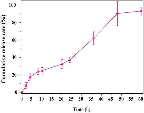
Figure 4 The anti-MRSA activity of GA/WS-CQDs hydrogels. Inhibition-zone test for 10 μg/mL penicillin hydrogels, 10 μg/mL vancomycin hydrogels, GA-gel, and GA/WS-CQDs-gel against MRSA, (A) Representative digital images and (B) statistical analysis showed antibacterial capacity by inhibition-zone test against MRSA (ATCC 43300). (C) Representative digital images and (D) statistical analysis against MRSA (22,212,587). The data are mean ± SD (n=3, statistical significance was analyzed via unpaired t-test, **p < 0.01, ****p < 0.001).
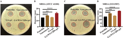
Figure 5 The biosafety assessment of GA/WS-CQDs hydrogels in vitro and in vivo. (A) Calcein-AM/PI staining images of cells treated with the exudate of different concentrations hydrogels for 24 h. Green fluorescence (live cells), red fluorescence (death cells). (B) Hemolysis assay. Saline and ddH2O as negative and positive control group, respectively. (C) The change of white blood cells (WBC), red blood cells (RBC), platelets (PLT), hemoglobin (HGB), ALT (alanine aminotransferase), LDH (lactate dehydrogenase), CK-MB (creatine kinase MB), UERA. ns represents not significant. (D) H&E staining images of heart, liver, spleen, lung, and kidney sections from mice with wounds. Scale bar, 100 μm.
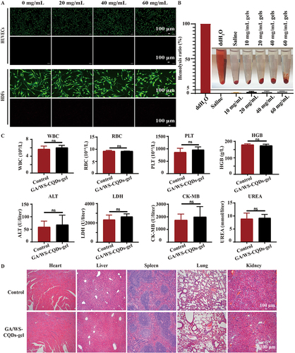
Figure 6 GA/WS-CQDs dressings accelerated MRSA-infected wounds healing in vivo. (A) Image of GA/WS-CQDs dressings. (B) Pattern diagram of MRSA-infected wound model and treatment. (C) Representative images of wounds treated with 3M film, GA dressings, and GA/WS-CQDs dressings at the indicated time points. Scale bar, 20 μm. (D) Statistical of the healing times in every group. **p < 0.01, *p < 0.05. (E) H&E and Masson’s trichrome staining of wounds skin tissue. Black dashed line, epidermis. Scale bar, 200 μm.
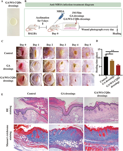
Figure 7 GA/WS-CQDs dressing inhibited wound inflammation in vivo. (A) Immunohistochemical staining images of proinflammatory factors including TNF-α, IL-1β and IL-6 in MRSA-infected wound skin tissues. Scale bar, 50 μm. (B) Quantification of the mean intensities for the areas positive from (A). ****p < 0.001, ***p < 0.005, **p < 0.01, and *p < 0.05.
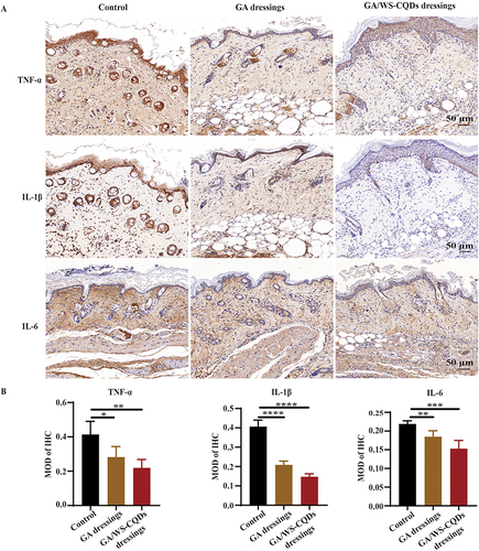
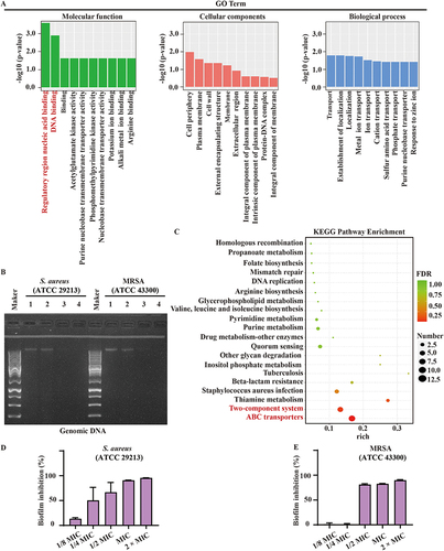
Figure 8 The antibacterial mechanism of WS-CQDs. RNA-Seq gene expression profiles. Control vs WS-CQDs treated groups. Red text indicates gene or pathways with the most significant differential expression. (A) Go enrichment analysis in molecular function (MF), cellular component (CC) and biological process (BP). (B) Gel electrophoretic mobility of WS-CQDs binding to bacteria genomic DNA. Band 1 (control), band 2 (1:0.3), band 3 (1:0.6) and band 4 (1:1.2). (C) KEGG enrmichent analysis. (D and E) Inhibition rates of the biofilm formation of S. aureus and MRSA.