
Figures & data
Figure 1 The process of preparing the nanoprobe.
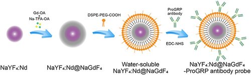
Figure 2 Characteristics of the NaYF4:Nd@NaGdF4-ProGRP antibody nanoprobe. (a) electron microscope image of NaYF4:Nd@NaGdF4 nanocrystals; (b) size of the NaYF4:Nd@NaGdF4-ProGRP antibody nanoprobe (44 nm); (c) emission spectrum of NaYF4:Nd@NaGdF4; (d) a near-infrared photograph of NaYF4:Nd@NaGdF4.
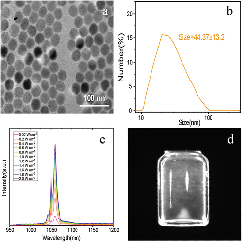
Figure 3 Toxicity of the NaYF4:Nd@NaGdF4-ProGRP antibody probe against HTB-119 cell proliferation (C, concentration of the probe).
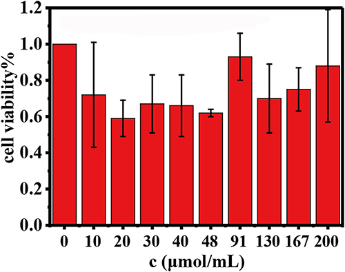
Figure 4 Near-infrared identification imaging with the probe. (a) micrograph of small-cell lung cancer HTB-119 cells; (b) DAPI imaging of small-cell lung cancer HTB-119 cells; (c) near-infrared identification imaging for HTB-119 cells with the NaYF4:Nd@NaGdF4-ProGRP antibody probe (cyan pseudocolor); (d) merged image of the HTB-119 cells identified by the NaYF4:Nd@NaGdF4-ProGRP antibody probe and those identified by DAPI imaging).
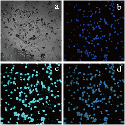
Figure 5 Nude mouse subcutaneous small-cell lung cancer tumor model. (a) BALB/c nude mouse; (b) a tumor-bearing nude mouse (the white arrow indicates the location of the subcutaneous tumor); (c) CT scan of the nude mouse lung cancer subcutaneous tumor model (the black arrow indicates the location of the subcutaneous tumor).
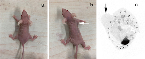
Figure 6 Near-infrared fluorescence images of the small-cell lung cancer HTB-119 subcutaneous tumor model in nude mice (the dashed line shows the best near-infrared fluorescence image, which was obtained 10 minutes after probe injection).
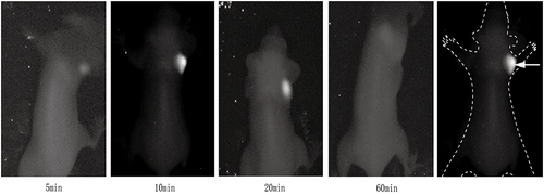
Figure 7 Image of HE-stained tissue area excised after identification of the subcutaneous small-cell lung cancer tumor in the nude mouse model with the probe (10 μm, the white arrow points to the cancer cells).
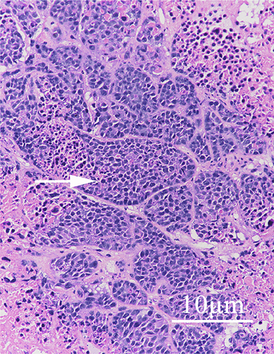
Table 1 Hematology, Hepatotoxicity and Nephrotoxicity of the Probe to Small Cell Lung Cancer HTB-119 Subcutaneous Tumor Model Mice