Figures & data
Figure 1 Schematic illustration of the green synthesis of silver nanoparticles using an aqueous extract of the Chrysanthemum indicum (flower).
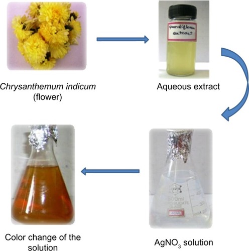
Table 1 Phytochemical screening of the flower extract of Chrysanthemum indicum
Figure 2 Ultraviolet–visible spectroscopy analysis of synthesized silver nanoparticles, and the peak noted around 435 nm.
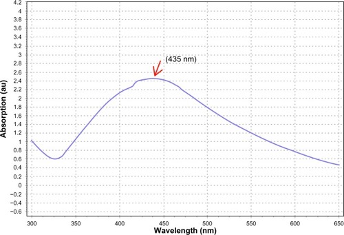
Figure 3 X-ray diffraction pattern of synthesized silver nanoparticles from the floral extract of Chrysanthemum indicum.
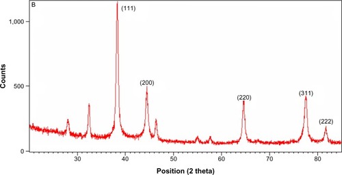
Figure 4 Transmission electron microscopy micrograph of the green synthesized silver nanoparticles.
Notes: Scale bars: (A) 200 nm and (B) 500 nm. The arrows indicate the particle size in nm.
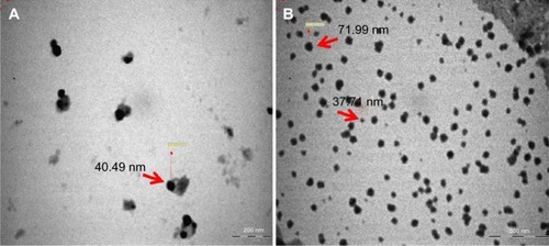
Figure 5 Energy-dispersive X-ray spectrum of green synthesized silver nanoparticles.
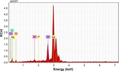
Table 2 Antibacterial activity of AgNPs synthesized by the aqueous flower extract of Chrysanthemum indicum
Figure 6 The antimicrobial efficacy of the aqueous extracts of Chrysanthemum indicum, AgNO3, and AgNPs were examined against three selected Gram-positive bacterial strains (D, E, and F) and three selected Gram-negative bacterial strains (A, B, and C).
Notes: The Gram-positive bacterial strains included BS (MTCC 121), SE (MTCC 435), and SA (MTCC 96), and the three Gram-negative bacterial strains included KP (MTCC 109), EC (MTCC 433), and PA (MTCC 1934). The arrows indicate the zone of inhibition.
Abbreviations: PE, plant extract; C, control (streptomycin); KP, Klebsiella pneumoniae; AgNP, silver nanoparticle; EC, Escherichia coli; PA, Pseudomonas aeruginosa; BS, Bacillus subtilis; SE, Staphylococcus epidermidis; SA, Staphylococcus aureus; MTCC, Microbial Type Culture Collection and Gene Bank.
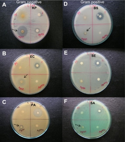
Table 3 Minimum inhibitory concentration of AgNPs synthesized by aqueous flower extract of Chrysanthemum indicum
Table 4 Cell viability and LDH leakage in control- and AgNP-treated 3T3 cells after 24 hours of exposure
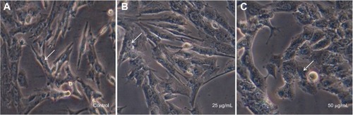
Figure 7 Cytotoxicity of synthesized silver nanoparticles on 3T3 mouse embryo fibroblast cells.
Notes: (A) Control (cell + phosphate buffered saline); (B) cell +25 μg/mL of silver nanoparticles; (C) cell +50 μg/mL of silver nanoparticles. The arrows indicate the cell nucleus.