Figures & data
Figure 1 Amino acid sequences of gB498–505 and gD301–309 peptide epitopes, the schematic representation of the corresponding peptide amphiphilic derivatives and of (C18)2-L adjuvant, and their molecular weights (Mw).
Note: The amino acid sequences are reported using the three-letter abbreviation.
Abbreviations: AdOO, 8-amino-3,6-dioxaoctanoic acid; Ala, alanine; Arg, arginine; Asp, aspartate; Glu, glutamic acid; Gly, glycine; Ile, isoleucine; Leu, leucine; Phe, phenylalanine; Pro, proline, Ser, serine; Val, valine.
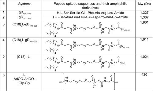
Table 1 List of the investigated aggregated systems, with the ratio (R) between the amphiphilic monomers, and their structural properties
Figure 2 Structural characterization of the aggregates, by fluorescence and dynamic light scattering.
Notes: (A) Fluorescence intensity of the ANS fluorophore at 480 nm as a function of PAs concentration: (C18)2-L-gB498–505 (□), (C18)2-L-gD301–309 (▲), and (C18)2-L-gB498–505/(C18)2-L-gD301–309 (•). CMC values were established from the graphical break point; (B) DLS spectra of (C18)2-L-gB498–505/(C18)2-L-gD301–309 mixed aggregates at 25°C and 1 × 10⁻⁴ M concentration.
Abbreviations: ANS, 8-anilino-1-naphthalene sulfonic acid ammonium salt; CMC, critical micelle concentration; DLS, dynamic light scattering; PA, peptide amphiphile.
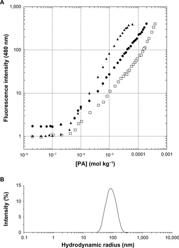
Figure 3 Far-UV CD spectra of: (A) gD301–309 peptide at 0, 40%, and 80% of TFE in TRIS buffer solution, after subtraction of gD301–309 spectrum at 0% TFE; similar spectra were observed for the gB498–505 peptide; (B) pure and mixed (50/50) amphiphilic epitopes at 2 × 10⁻⁴ M concentration, well above the CMC values to ensure the presence of aggregates in solution.
Abbreviations: CMC, critical micelle concentration; MRE, mean residue ellipticity; TFE, trifluoroethanol; TRIS, tris(hydroxymethyl)aminomethane; UV-CD, ultraviolet circular dichroism.
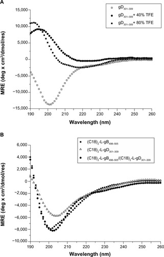
Figure 4 Effects of PAs (gB, gD, gB/gD) and PA-micelles on cell viability.
Notes: Cell viability was evaluated by MTT assay at 24 hours posttreatment in U937 (A) and RAW 264.7 macrophages (B). Cisplatin (20 μM) was used as a positive control of cell death. Values are expressed as the mean ± SD of three independent experiments.
Abbreviations: MTT, 3-(4,5-dimethylthiazol-2-yl)-2,5-diphenyltetrazolium bromide; PA, peptide amphiphile; SD, standard deviation.
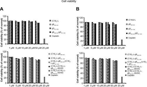
Figure 5 IL-17- and IL-23-release after treatment with 10 μM of each peptide and peptide-micelles for 24 hours.
Notes: LPS was used as a positive control. The results shown are the average of three independent experiments, and the error bars indicate the standard errors of the means. *P≤0.01 indicates statistically significant difference between untreated cells versus single/mixed peptide pretreated cells (Student’s t-test).
Abbreviations: IL, interleukin; LPS, lipopolysaccharide.
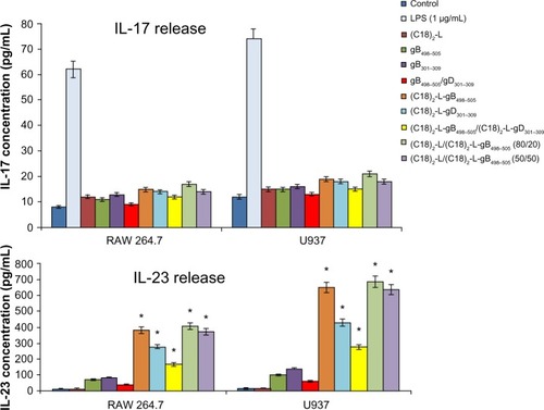
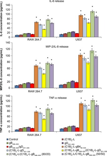
Figure 6 IL-6-, IL-8- or MIP-2-, and TNF-α-release after treatment with 10 μM of each peptide and peptide-micelles, for 24 hours.
Notes: The results shown are the average of three independent experiments, and the error bars indicate the standard errors of the means. *P≤0.01 indicates a statistically significant difference between untreated cells versus single/mixed peptides pretreated cells (Student’s t-test).
Abbreviations: IL, interleukin; MIP, macrophage inflammatory protein; TNF, tumor necrosis factor.