Figures & data
Figure 1 Chemical structures of antifungals used in this study. (A) Amphotericin B; (B) natamycin; (C) fluconazole; (D) itraconazole; (E) terbinafine chloride.
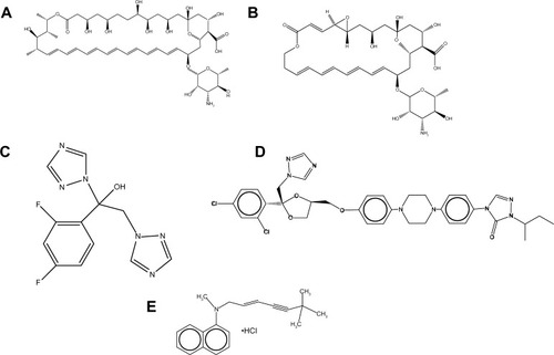
Figure 2 Scanning electron micrographs showing the electrospun gelatin fibers loaded with various antifungals. (A) No antifungals; (B) amphotericin B; (C) natamycin; (D) fluconazole; (E) itraconazole; (F) terbinafine. (G) Histogram showing the effects of various antifungals on the average fiber diameter.
Notes: ***P<0.001; scale bar =50 μm.
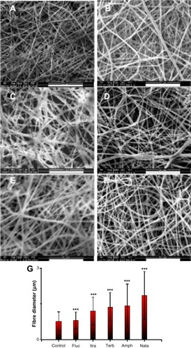
Figure 3 Efficacy of drug-loaded gelatin fiber mats against yeast strains. The antifungal activity against Candida albicans strains is expressed as the zone of inhibition measured by radial diffusion assay. Fiber mats loaded with (A) amphotericin B (*P<0.05 compared to C. albicans ATCC 10231 strains; **P<0.01 compared to C. albicans ATCC 10231 strains; @@P<0.01 compared to C. albicans ATCC 2091 strains; @@@P<0.001 compared to C. albicans ATCC 2091 strains; #P<0.05 compared to C. albicans ATCC 24433); (B) natamycin (*P<0.05 compared to C. albicans ATCC 10231 strains); (C) terbinafine (***P<0.001 compared to all the groups); (D) itraconazole (***P<0.01 compared to all the groups except C. albicans ATCC 24433 strains; @@@P<0.001 compared to all the groups except C. albicans ATCC 2091 strains); and (E) fluconazole. ***P<0.001 compared to all the groups. (F) Representative photographs showing the zone of inhibition of gelatin (upper panel) and amphotericin B-loaded gelatin (lower panels) fiber mats.
Note: The absence of bars in the graph indicates no inhibition against the particular strain.
Abbreviation: ATCC, American Tissue Culture Collection.
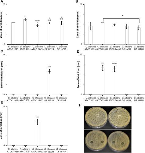
Figure 4 Efficacy of drug-loaded gelatin fiber mats against filamentous fungi. The antifungal activity against Aspergillus and Fusarium strains is expressed as the zone of inhibition measured by radial diffusion assay. Fiber mats loaded with (A) amphotericin B (***P<0.001 compared to Aspergillus brasiliensis ATCC 16404); (B) natamycin (*P<0.05 compared to Fusarium solani ATCC 3636 strains; @P<0.05 compared to A. brasiliensis ATCC 16404 strains); (C) terbinafine (***P<0.001 compared to F. solani ATCC 3636 strains; @@@P<0.001 compared to A. brasiliensis ATCC 16404); (D) itraconazole (***P<0.001 compared to F. solani ATCC 3636 strains; @@@P<0.001 compared to F. solani ATCC 26671 strains; #P<0.05 compared to A. fumigatus ATCC 90906 strains); and (E) fluconazole. (F) Representative photographs showing the zone of inhibition of gelatin (upper panel) and amphotericin B-loaded gelatin (lower panels) fiber mats.
Note: The absence of bars in the graph indicates no inhibition against the particular strain.
Abbreviation: ATCC, American Tissue Culture Collection.
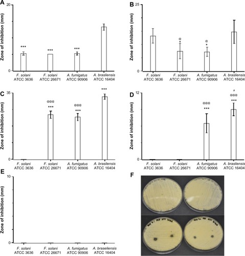
Table 1 Minimum inhibitory concentration of antifungals against various strains of Candida albicans
Figure 5 Cumulative release profile of antifungals form gelatin fiber mats.
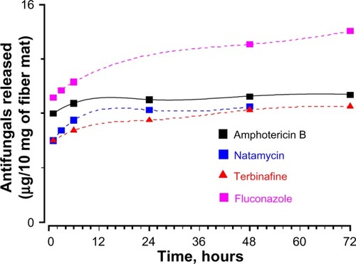
Figure 6 (A) Candidacidal properties of the fiber mats loaded with amphotericin B and natamycin. (B) Kinetics of candidacidal action of amphotericin B and natamycin-loaded fiber mats.
Notes: There was a significant decrease in the viability of Candida albicans exposed to antifungal-loaded fiber mats; **P<0.01.
Abbreviation: AmpB, amphotericin B.
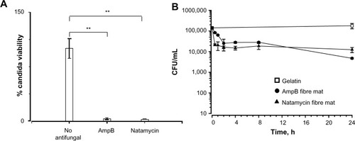
Figure 7 Morphology of Candida albicans grown on polyene-loaded fiber mats. Scanning electron micrographs of C. albicans treated with (A) gelatin fiber mats without antifungals; (B) amphotericin B-loaded fiber mats; and (C) natamycin-loaded fiber mats.
Note: Inset scale bar =10 μm.
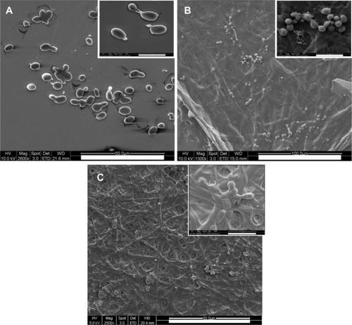
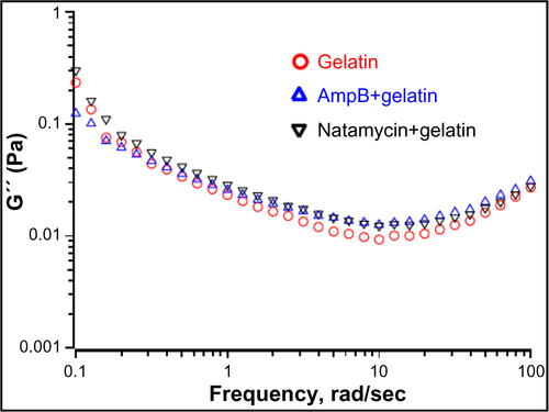
Figure 8 Interaction between polyene antifungals and gelatin probed by circular dichroism spectropolarimetry and rheological studies. (A) Far ultraviolet circular dichroism spectropolarimetry of gelatin and gelatin incubated with polyene antifungals at 30°C. Thermal denaturation of gelatin in the presence of (B) amphotericin B and (C) natamycin. The concentration of antifungals was 0.25 wt% with respect to gelatin. (D) Frequency sweep of storage modulus (G′) of gelatin and antifungal-loaded gelatin.
Abbreviation: AmpB, amphotericin B.
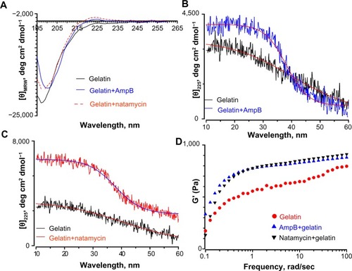
Figure 9 (A) Ultraviolet absorption spectra of amphotericin B in three different media. The four peaks observed in the monomeric amphotericin B are labelled. The concentration of amphotericin used was 50 μg/mL. The Y-axis for the amphotericin B in gelatin is represented separately. (B) The relative aggregation state of amphotericin B as measured by the intensity ratio of peak I and peak IV of the absorption spectra in three different media. (C) Circular dichroism spectrum of amphotericin B in three different media. Note the bisignate nature of amphotericin B in phosphate buffered saline indicating a complex structure. (D) Hemolytic activity of amphotericin B and natamycin in the presence/absence of gelatin. For all these studies, the antifungal:gelatin ratio was 0.25 wt%. (E) Ultraviolet absorption spectra of amphotericin B released from the gelatin matrix. For a comparison, the absorption spectra of free amphotericin B (10 μg/mL) in buffer is also shown.
Note: ***P<0.001.
Abbreviations: AmpB, amphotericin B; DMSO, dimethyl sulfate; PBS, phosphate buffered saline; AU, arbitrary unit.
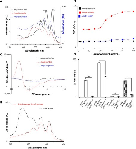
Figure 10 Cytotoxicity of polyene-loaded antifungal fiber mats against (A) human corneal and (B) human sclera fibroblasts. XL-gelatin fiber mat is the gelatin crosslinked with glutaraldehyde for 24 hours. For amphotericin B- and natamycin-loaded fiber mats the crosslinking time was 3 hours.
Notes: *P<0.05 compared to XL-gelatin fiber mats; **P<0.01 compared to XL-gelatin fiber mats; @P<0.05 compared to control.
Abbreviation: AmpB, amphotericin B.
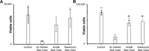
Table 2 Mechanical properties of polyene-loaded fiber mats
Figure 11 Mechanical properties of glutaraldehyde vapor crosslinked gelatin nanofibers with and without drug loading. The gelatin fiber mat was crosslinked with glutaraldehyde for 48 hours. For amphotericin B- and natamycin-loaded gelatin fiber mats 3-hour glutaraldehyde crosslinking was used.
Abbreviation: AmpB, amphotericin B.
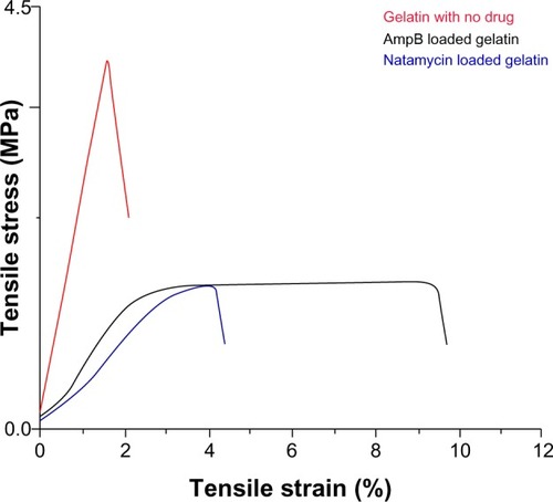
Figure S1 Growth of yeast/fungal pathogens on Sabouraud dextrose agar plate.
Abbreviations: C. albicans, Candida albicans; F. solani, Fusarium solani; A. brasiliensis, Aspergillus brasiliensis; A. fumigatus, Aspergillus fumigatus.
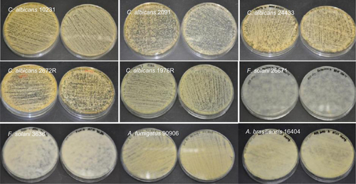
Figure S2 Growth of yeast/fungal pathogens on gelatin fiber mats (without any antifungals).
Abbreviations: C. albicans, Candida albicans; F. solani, Fusarium solani; A. brasiliensis, Aspergillus brasiliensis; A. fumigatus, Aspergillus fumigatus.
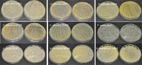
Figure S3 Antifungal properties of amphotericin B-loaded electrospun fiber mats.
Abbreviations: C. albicans, Candida albicans; F. solani, Fusarium solani; A. brasiliensis, Aspergillus brasiliensis; A. fumigatus, Aspergillus fumigatus.
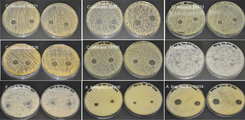
Figure S4 Antifungal properties of natamycin-loaded electrospun fiber mats.
Abbreviations: C. albicans, Candida albicans; F. solani, Fusarium solani; A. brasiliensis, Aspergillus brasiliensis; A. fumigatus, Aspergillus fumigatus.
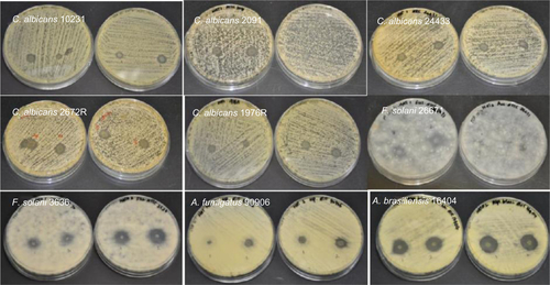
Figure S5 Antifungal properties of fluconazole-loaded electrospun fiber mats.
Abbreviations: C. albicans, Candida albicans; F. solani, Fusarium solani; A. brasiliensis, Aspergillus brasiliensis; A. fumigatus, Aspergillus fumigatus.
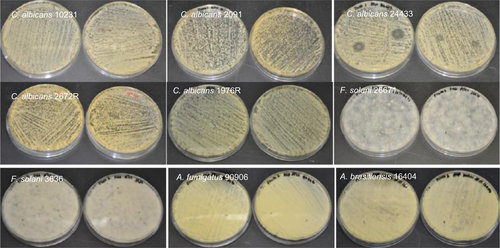
Figure S6 Antifungal properties of itraconazole-loaded electrospun fiber mats. In some strains of C. albicans a weak inhibition was observed.
Abbreviations: C. albicans, Candida albicans; F. solani, Fusarium solani; A. brasiliensis, Aspergillus brasiliensis; A. fumigatus, Aspergillus fumigatus.
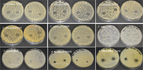
Figure S7 Antifungal properties of terbinafine-loaded electrospun fiber mats.
Abbreviations: C. albicans, Candida albicans; F. solani, Fusarium solani; A. brasiliensis, Aspergillus brasiliensis; A. fumigatus, Aspergillus fumigatus.
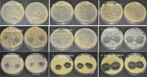
Figure S8 (A) Far ultraviolet circular dichroism spectropolarimetry of gelatin and gelatin incubated with fluconazole and terbinafine at 30°C. Thermal denaturation of gelatin monitored by changes in (θ)225 in the presence of (B) fluconazole and (C) terbinafine. The concentration of antifungals was 0.25 wt% with respect to gelatin.
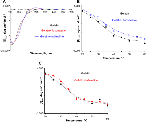
Figure S9 Frequency sweep showing the effect of polyenes on loss modulus (G″).
Abbreviation: AmpB, amphotericin B.