Figures & data
Table 1 Structural characteristics of void and ZnPc containing liposomes. Values given are mean values of three different batches ± standard deviation of the mean
Figure 1 Uptake of Rh-PE-labeled liposomes by J774 cells as a function of time.
Notes: J774 cells were incubated with Rh-PE-Ls, Rh-PE-UDLs, Rh-PE-ARCs, and Rh-PE-UDAs at 0.5 mM phospholipids in medium containing 5% FCS. At different time points, cells were washed with phosphate-buffered saline, collected, fixed, and analyzed by flow cytometry (BD FACSCalibur™; BD Biosciences, San Jose, CA, USA).
Abbreviations: Rh-PE, dimyristoyl phosphoethanolamine-N-(Lissamine rhodamine B sulfonyl); Ls, conventional liposomes; UDLs, ultradeformable liposomes; ARCs, archaeosomes; UDAs, ultradeformable archaeosomes; FCS, fetal calf serum.
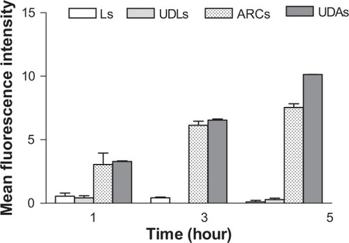
Figure 2 Cytotoxicity of free and liposomal ZnPc on mammal cells in the darkness and after irradiation.
Notes: HaCaT cells (A), J774 cells (B), and dendritic cells (C) were incubated with 0.01 μM and 0.1 μM of ZnPc, ZnPcUDLs, and ZnPcALs for 24 hours in medium containing 5% FCS. Half the cells were kept in the darkness, and half were irradiated. After irradiation, cells were incubated for 24 hours in growth medium. Cell survival was determined by the 3-(4,5-dimethylthiazol-2-yl)-2,5-diphenyltetrazolium bromide assay. Values represent means ± standard deviation (n=3). **P<0.01; *P<0.05.
Abbreviations: ZnPc, zinc phthalocyanine; ZnPcUDLs, ZnPc containing ultradeformable liposomes; ZnPcALs, ZnPc and archaeolipids containing liposomes; FCS, fetal calf serum.
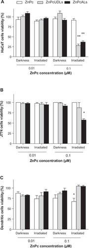
Figure 3 Cytotoxicity of free and liposomal ZnPc on Leishmania braziliensis promastigotes in the darkness and after irradiation.
Notes: L. braziliensis promastigotes were incubated with 0.01 μM and 0.1 μM of ZnPc, ZnPcUDLs, and ZnPcALs for 24 hours in Schneider’s Insect Medium (Sigma Aldrich, St Louis, MO, USA) at 26°C. Half the parasites were kept in darkness, and half were irradiated. After irradiation, parasites were incubated for 24 hours in growth medium. Cell survival was determined by the 3-(4,5-dimethylthiazol-2-yl)-2,5-diphenyltetrazolium bromide assay. Values represent means ± standard deviation (n=3).
Abbreviations: ZnPc, zinc phthalocyanine; ZnPcUDLs, ZnPc containing ultradeformable liposomes; ZnPcALs, ZnPc and archaeolipids containing liposomes.
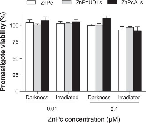
Figure 4 Antiamastigote activity of free or liposomal ZnPc in the darkness and after irradiation.
Notes: (A) J774 cells previously infected with Leishmania braziliensis promastigotes were incubated with 0.01 μM (I) and 0.1 μM (II) of ZnPc, ZnPcUDLs, and ZnPcALs, and with void liposomes (UDLs and ALs at the phospholipid concentrations 7.6–5.2 [I] μM and 76–52 [II] μM) for 4 hours in medium containing 5% fetal calf serum. Half the cells were kept in darkness, and half were irradiated. After irradiation, cells were incubated for 24 hours in growth medium. Then cells were fixed with methanol and stained with Giemsa (Merck, New Jersey, USA). The number of amastigotes/100 cells was determined by counting at least 300 cells in three different experiments, and antiamastigote activity was calculated. (B,C) Optical microscopy of infected J774 cells incubated with 0.01 μM ZnPc (B) and 0.01 μM ZnPcALs (C), both taken 24 hours after irradiation. Arrows points to intracellular amastigotes. *P<0.05; ***P<0.001.
Abbreviations: ZnPc, zinc phthalocyanine; ZnPcUDLs, ZnPc containing ultradeformable liposomes; ZnPcALs, ZnPc and archaeolipids containing liposomes; UDLs, ultradeformable liposomes; ALs, archaeolipids containing liposomes.
![Figure 4 Antiamastigote activity of free or liposomal ZnPc in the darkness and after irradiation.Notes: (A) J774 cells previously infected with Leishmania braziliensis promastigotes were incubated with 0.01 μM (I) and 0.1 μM (II) of ZnPc, ZnPcUDLs, and ZnPcALs, and with void liposomes (UDLs and ALs at the phospholipid concentrations 7.6–5.2 [I] μM and 76–52 [II] μM) for 4 hours in medium containing 5% fetal calf serum. Half the cells were kept in darkness, and half were irradiated. After irradiation, cells were incubated for 24 hours in growth medium. Then cells were fixed with methanol and stained with Giemsa (Merck, New Jersey, USA). The number of amastigotes/100 cells was determined by counting at least 300 cells in three different experiments, and antiamastigote activity was calculated. (B,C) Optical microscopy of infected J774 cells incubated with 0.01 μM ZnPc (B) and 0.01 μM ZnPcALs (C), both taken 24 hours after irradiation. Arrows points to intracellular amastigotes. *P<0.05; ***P<0.001.Abbreviations: ZnPc, zinc phthalocyanine; ZnPcUDLs, ZnPc containing ultradeformable liposomes; ZnPcALs, ZnPc and archaeolipids containing liposomes; UDLs, ultradeformable liposomes; ALs, archaeolipids containing liposomes.](/cms/asset/8722531d-96cd-43ba-98bc-b11a671bc8a0/dijn_a_60543_f0004_c.jpg)
Figure 5 Uptake of liposomal ZnPc by J774 cells.
Notes: (A) J774 cells were incubated with 0.01 μM of ZnPc, ZnPcUDLs, and ZnPcALs for 24 hours in medium containing 5% fetal calf serum (FCS). Then, cells were washed with phosphate-buffered saline (PBS), suspended in PBS, and analyzed by flow cytometry (BD FACSCalibur™; BD Biosciences, San Jose, CA, USA). (B) J774 cells were preincubated with 4 mg/mL mannan for 30 minutes in medium containing 5% FCS, then 0.1 μM of ZnPcUDLs and ZnPcALs were added and incubated for another hour at 37°C. Then, cells were washed with PBS, suspended in PBS, and analyzed by flow cytometry. Values represent mean ± standard deviation (n=3). ***P<0.001.
Abbreviations: ZnPc, zinc phthalocyanine; ZnPcUDLs, ZnPc containing ultradeformable liposomes; ZnPcALs, ZnPc and archaeolipids containing liposomes; NS, not significant.
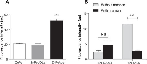
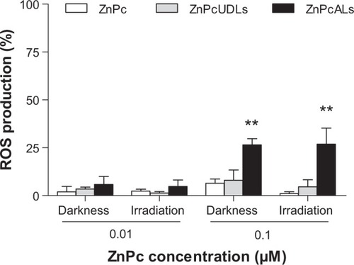
Figure 6 Reactive oxygen species (ROS) production of J774 cells after treatment with free or liposomal ZnPc in darkness and after irradiation.
Notes: J774 cells were incubated with 0.01 μM and 0.1 μM of ZnPc, ZnPcUDLs, and ZnPcALs for 4 hours in medium containing 5% fetal calf serum. Half the cells were kept in darkness, and half were irradiated. After irradiation, cells were incubated for 30 minutes at 37°C, and then medium was removed and replaced by 10 μM of (5-and-6)-chloromethyl-2′,7′-dichlorodihydrofluorescein diacetate, acetyl ester in phosphate-buffered saline (PBS). Then, cells were washed with PBS, suspended in PBS, and analyzed by flow cytometry (BD FACSCalibur™; BD Biosciences, San Jose, CA, USA). Values represent means ± standard deviation (n=3). **P<0.01.
Abbreviations: ZnPc, zinc phthalocyanine; ZnPcUDLs, ZnPc containing ultradeformable liposomes; ZnPcALs, ZnPc and archaeolipids containing liposomes.