Figures & data
Table 1 The dosage of DOX for pharmacodynamic test
Figure 1 A comparison of the solubilities of DOX in different salt solutions as a function of pH. The solubilities of DOX were quantified after a 24-hour incubation of 2 mg DOX within 1 mL of 200 mM salt solutions of different pH at 25°C.
Note: **P<0.01 versus (NH4)2SO4 group.
Abbreviations: DOX, doxorubicin; EDTA, ethylenediaminetetraacetic acid.
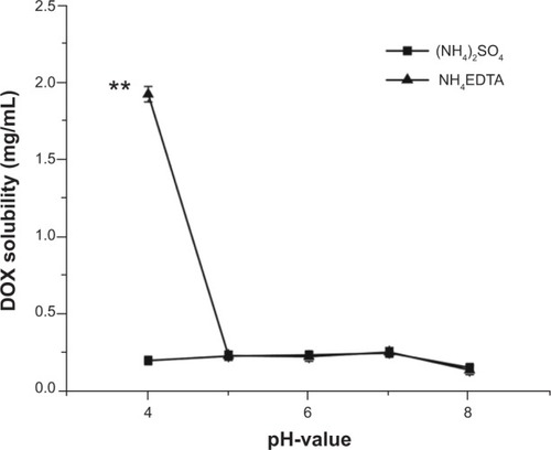
Figure 2 Photographs of mixtures of DOX with (1) (NH4)2SO4 and (2) NH4EDTA at 25°C and pH 4 (A) before centrifugation, (B) after centrifugation, and (C) diluted with isochoric distilled water.
Abbreviations: DOX, doxorubicin; EDTA, ethylenediaminetetraacetic acid.
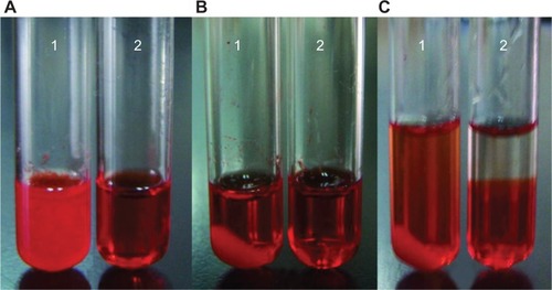
Figure 3 A comparison of the release of DOX encapsulated in liposomes in vitro via different ion gradients: 200 mM NH4EDTA solution and 200 mM (NH4)2SO4 solutions. Drug release profiles were analyzed in conventional PBS buffer (pH 7.4) and a special PBS buffer with ammonium chloride (80 mM), histidine (10 mM), penicillin (100 μg/mL), and streptomycin (100 μg/mL) (pH 7.4).
Note: **P<0.01 versus (NH4)2SO4-L in PBS buffer with ammonium chloride.
Abbreviations: DOX, doxorubicin; EDTA, ethylenediaminetetraacetic acid; L, doxorubicin-loaded liposomes; PBS, phosphate-buffered saline.
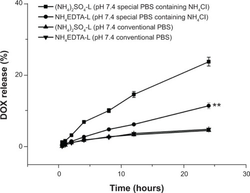
Table 2 Long-term stability of HSPC/CH/mPEG2000-DSPE (3/1/1, w/w/w) liposomes loaded with doxorubicin (drug/lipid ratio 1:10, w/w) via 200 mM NH4EDTA and (NH4)2SO4 gradient stored at 4°C±2°C
Figure 4 (A) Tumor inhibition effects of different DOX formulations. The drug doses were 5.0 mg/kg for DOX solution and (NH4)2SO4-L, and 2.5, 5.0, 10.0 mg/kg for the low, middle, and high concentration of NH4EDTA-L, respectively. (B) Damage of different formulations on immune organs of H22-bearing mice.
Notes: The data represent mean ± standard deviation. *P<0.05 versus NS-treated mice; **P<0.01 versus NS-treated mice; #P<0.05 versus DOX solution-treated mice.
Abbreviations: DOX, doxorubicin; EDTA, ethylenediaminetetraacetic acid; L, doxorubicin-loaded liposomes; NS, normal saline.
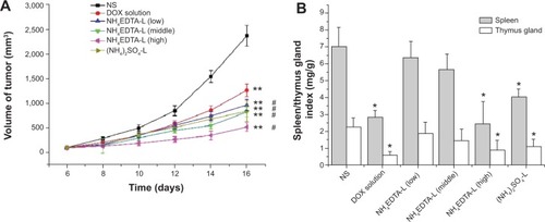
Figure 5 Tissue distributions of DOX in mice after intravenous administration of (A) 5 mg/kg DOX solution, NH4EDTA-L, and (NH4)2SO4-L and (B) 5.0 mg/kg DOX solution and different doses of NH4EDTA-L (2.5, 5.0, and 10.0 mg/kg).
Notes: The data represent mean ± standard deviation (n=10). Differences between the groups versus DOX solution are statistically significant at the P<0.01 level.
Abbreviations: DOX, doxorubicin; EDTA, ethylenediaminetetraacetic acid; L, doxorubicin-loaded liposomes.
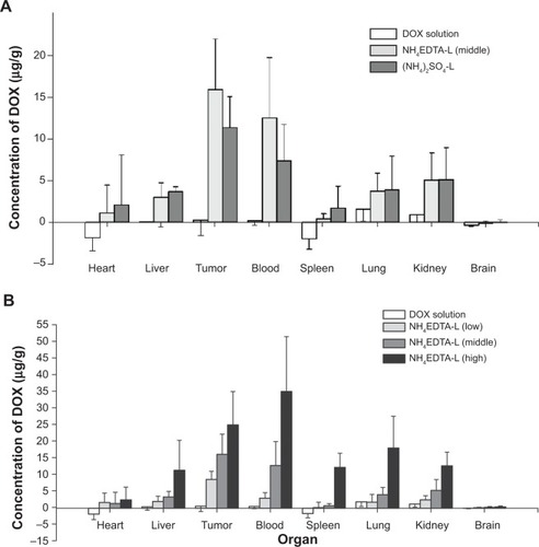
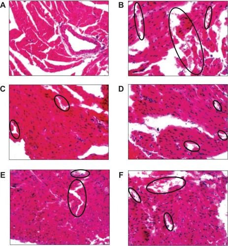
Figure 6 Typical images of pathological classification of myocardium injury of different DOX formulations: (A) NS; (B) DOX solution; (C) NH4EDTA-L (low-dose); (D) NH4EDTA-L (medium-dose); (E) NH4EDTA-L (high-dose); and (F) (NH4)2SO4-L.
Notes: Photographs were obtained under a fluorescence microscope (IX71; Olympus Corporation, Tokyo, Japan) using a 40× objective. The black circles point out the damaged sites of myocardium fragmentation.
Abbreviations: DOX, doxorubicin; EDTA, ethylenediaminetetraacetic acid; L, doxorubicin-loaded liposomes; NS, normal saline.