Figures & data
Figure 1 Preparation of BF/Gr and LV/Gr dispersions and representation of possible conformations for Gr in the DODAB bilayer: dimeric channel conformation in LV and intertwined Gr molecules at the borders of BF.
Abbreviations: DODAB, dioctadecyldimethylammonium bromide; LV, closed bilayers; BF, bilayer disks; Gr, Gramicidin; TFE, 2,2,2-trifluoroethanol.
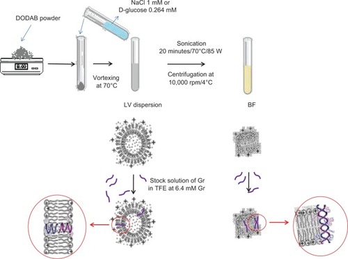
Table 1 Physical characterization of DODAB BF or LV, DODAB BF/Gr, or DODAB LV/Gr dispersions prepared in a 1 mM solution of NaCl
Table 2 DODAB or Gr concentrations (%) in BF/Gr or LV/Gr dispersions before and after filtering
Figure 2 Gr intrinsic fluorescence at λexc =280 nm and slit widths of 2.5 nm in different types of medium (A). Gr circular dichroism spectra at 25°C in different types of medium (B). In both subfigures, the Gr concentration is 0.02 mM and the DODAB concentration is 0.2 mM.
Abbreviations: DODAB, dioctadecyldimethylammonium bromide; LV, closed bilayers; BF, bilayer disks; Gr, Gramicidin; TFE, 2,2,2-trifluoroethanol.
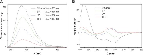
Figure 3 Effect of KCl or glucose concentration gradients across the LV on the normalized turbidity of the dispersions without (A–C) or with (D–F) Gr inserted in the DODAB LV. The Gr concentration is 0.1 mM and the DODAB concentration is 1.0 mM. The kinetics were obtained at 25°C.
Abbreviations: DODAB, dioctadecyldimethylammonium bromide; LV, closed bilayers; Gr, Gramicidin.
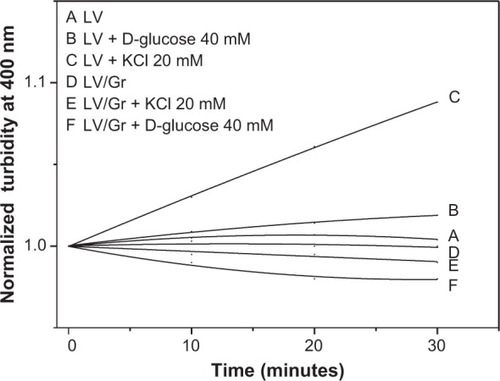
Figure 4 Cell viability (%) of Escherichia coli, Staphylococcus aureus, and Saccharomyces cerevisae as a function of Gr or DODAB. DODAB was dispersed as BF or LV, that were further added of Gr for a final DODAB to Gr molar ratio of about 10:1. The interaction time between the microbial cells and the dispersions was one hour, at final cell concentrations of 1.0×10⁸ CFU/mL (E. coli or S. aureus) or 1.0×10⁵ CFU/mL (S. cerevisae).
Abbreviations: CFU, colony-forming units; DODAB, dioctadecyldimethylammonium bromide; LV, closed bilayers; BF, bilayer disks; Gr, Gramicidin.
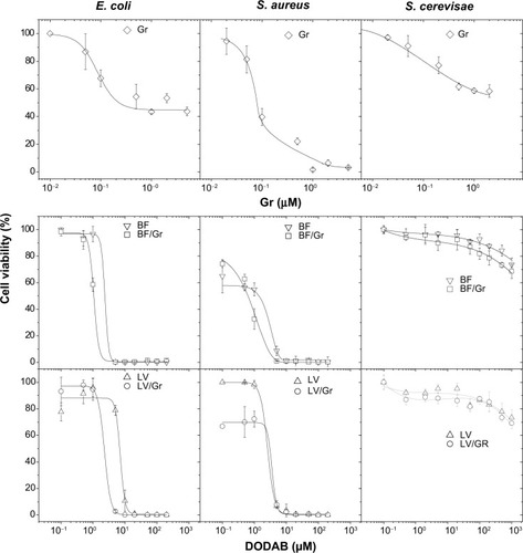
Table 3 Minimum bactericidal concentrations (in μM) and cell lysis at the minimum bactericidal concentration (% L) for DODAB, Gr, and DODAB/Gr dispersions
Figure 5 Leakage of phosphorylated compounds (%) from bacteria for different dispersions at 3.7×10⁸ to 4.8×10⁹ CFU/mL (Escherichia coli) or 5.2×10⁹ to 5.1×10¹⁰ CFU/mL (Staphylococcus aureus). The dispersions were tested over a range of DODAB or DODAB/Gr concentrations after one hour of interaction with both types of bacteria. As a control, over a range of Gr concentrations (10−2 to 10−4 mM), no leakage was detected from bacteria after the same interaction time.
Abbreviations: CFU, colony-forming units; DODAB, dioctadecyldimethylammonium bromide; LV, closed bilayers; BF, bilayer disks; Gr, Gramicidin.
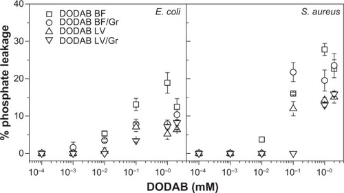
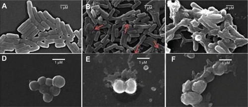
Figure 6 Micrographs of Escherichia coli (A–C) or Staphylococcus aureus (D–F) cells untreated (A, D) or treated with DODAB BF/Gr (B, E) or DODAB LV/Gr dispersions (C, F), obtained by scanning electron microscopy. Cells appear enlarged by 10,000× (E. coli) or 20,000× (S. aureus). Against E. coli cells, in (B), DODAB =0.005 mM (DODAB BF/Gr) and, in (C), DODAB =0.01 mM (DODAB LV/Gr). Against S. aureus, in (E), DODAB =0.01 mM (DODAB BF/Gr) and, in (F), DODAB =0.05 mM (DODAB LV/Gr).
Abbreviations: DODAB, dioctadecyldimethylammonium bromide; LV, closed bilayers; BF, bilayer disks; Gr, Gramicidin.