
Figures & data
Figure 1 Synthesis of gold nanoparticles (Au-NPs) by laser ablation. (A) Schematics of laser ablation in aqueous solution. (B) Extinction spectrum of Au-NPs prepared by femtosecond laser ablation and further fragmentation in deionized water. The inset shows an image of a typical solution of Au-NPs. Transmission electron microscopy images and corresponding size distributions of laser-synthesized Au-NPs in deionized water (C), Au-NPs in solution of polyethylene glycol (D), and Au-NPs in solution of dextran (E).
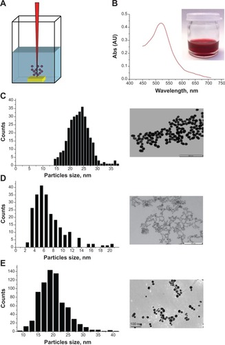
Table 1 Physicochemical characterization of Au-NPs
Figure 2 Uptake of Au-NPw, Au-NPp, and Au-NPd in human cancer cells. (A) Transmission electron microscopy images of neuroblastoma cell line cells incubated with 1 mg/L Au-NPw (a), Au-NPp (b), or Au-NPd (c) for 24 hours. (B) Intracellular trafficking of Au-NPw in neuroblastoma cell line cells. NPs are closed to the cell membrane (arrow) (a), enclosed in endocytic vesicles close to the cell membrane (arrow) (b), inside late endosome (arrow) (c), and inside endolysosome (full arrow) and lysosome (dotted arrow) (d). (C) Transmission electron microscopy images of glioblastoma cell line cells incubated with 1 mg/L Au-NPw for 24 hours. Au-NPw are internalized in glioblastoma cells (left) via endocytic vesicles (right).
Abbreviations: Au-NPs, gold nanoparticles; Au-NPd, Au-NPs prepared in dextran; Au-NPp, Au-NPs prepared in polyethylene glycol; Au-NPw, Au-NPs in pure deionized water.
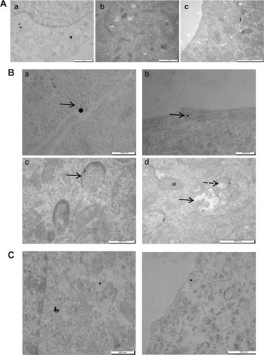
Figure 3 Safety of Au-NPs on human cancer cells. (A) MTT assay on SK-N-SH cells (left) and U87-MG cells (right) treated with Au-NPw, Au-NPp, and Au-NPd for 72 hours. (B) CellTiter-Glo® assay on SK-N-SH cells (left) and U87-MG cells (right) treated with Au-NPw for 72 hours. (C) MTT assay on SK-N-SH cells (left) and U87-MG cells (right) treated with paclitaxel as a positive control.
Abbreviations: Au-NPs, gold nanoparticles; Au-NPd, Au-NPs prepared in dextran; Au-NPp, Au-NPs prepared in polyethylene glycol; Au-NPw, Au-NPs in pure deionized water; MTT, 3-(4,5-dimethylthiazol-2-yl)-2,5-diphenyltetrazolium bromide; SK-N-SH, neuroblastoma; U87-MG, glioblastoma.

Figure 4 Morphology of cancer cells after Au-NP incubation for 6 hours at 1 mg/L. (A) Phase-contrast microscopy (10×) of neuroblastoma cells control (a) or treated with Au-NPw (b), Au-NPp (c), or Au-NPd (d) at 1 mg/L. (B) Immunofluorescence imaging of microtubular network in glioblastoma cells control (a), incubated for 6 hours with Au-NPw at 1 mg/L (b) or paclitaxel at 100 nM (c). Full arrow and dotted arrow show pseudoaster and bundles, respectively.
Abbreviations: Au-NPs, gold nanoparticles; Au-NPd, Au-NPs prepared in dextran; Au-NPp, Au-NPs prepared in polyethylene glycol; Au-NPw, Au-NPs in pure deionized water.
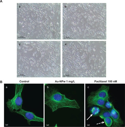
Figure 5 Time evolution and composition of protein corona. (A) Sodium dodecyl sulfate–polyacrylamide gel of corona protein on Au-NPw, Au-NPp, and Au-NPd at 30 minutes, 1 hour, and 24 hours. Left lane: protein molecular weight markers. (B) ImageJ profile of protein corona formed on each Au-NP at 30 minutes, 1 hour, and 24 hours. (C) Histograms representing the expression of C3 complement proteins and apolipoproteins (A-I, A-II, and E) adsorbed on Au-NPw, Au-NPp, and Au-NPd.
Abbreviations: Au-NPs, gold nanoparticles; Au-NPd, Au-NPs prepared in dextran; Au-NPp, Au-NPs prepared in polyethylene glycol; Au-NPw, Au-NPs in pure deionized water; emPAI, Exponentially Modified Protein Abundance Index.
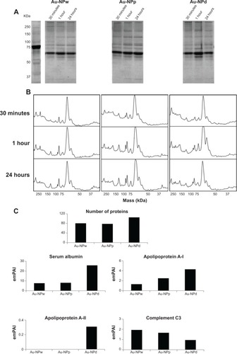
Figure S1 Cellular uptake of gold nanoparticles of different sizes in pure deionized water. Transmission electron microscopy images of neuroblastoma cells incubated with 1 mg/L gold nanoparticles in pure deionized water of 6 nm (A), 12 nm (B), or 19 nm (C) or polydispersed (D) for 24 hours.
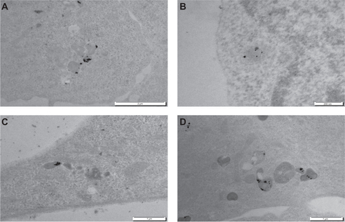
Table S1 List of the most abundant proteins identified on Au-NPw, Au-NPp, and Au-NPd by mass spectroscopy