Figures & data
Figure 1 Schematic diagram of Ag-MWCNTs’ preparation and evaluation of their antibacterial activity.
Abbreviation: MWCNT, multi-walled carbon nanotube.
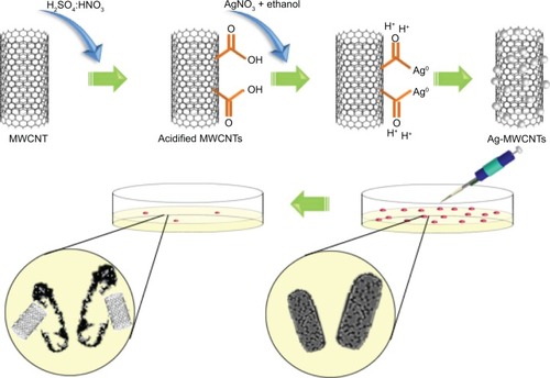
Figure 2 Physicochemical characterization of Ag-MWCNTs.
Notes: (A and B) TEM images of MWCNTs decorated with Ag nanoparticles. (C) EDS element map and pattern (yellow box is the area chosen for EPS analysis), and (D) EDS element analysis of Ag-MWCNTs. (E) XRD pattern of Ag-MWCNTs and Acidified MWCNTs. (F) Size distribution of Ag nanoparticles as assessed via TEM (n=100).
Abbreviations: CNT, carbon nanotube; EDS, energy-dispersive X-ray spectroscopy; MWCNT, multi-walled CNT; TEM, transmission electron microscopy; XRD, X-ray diffraction.
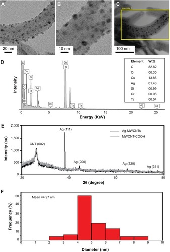
Figure 3 Antibacterial evaluation of Ag-MWCNTs.
Notes: (A) Antibacterial activity of different samples against Sphingomonas spp. and Methylobacterium spp. Concentration (CFU/mL): *P<0.05, one-tailed Mann–Whitney U-test. Data are representative of three experiments. (B) Evaluation of inhibition zones by different samples for Methylobacterium spp. and (C) Sphingomonas spp.
Abbreviations: CFU, colony forming units; MWCNT, multi-walled carbon nanotube, MWCNTs-COOH, acidified MWCNTs.
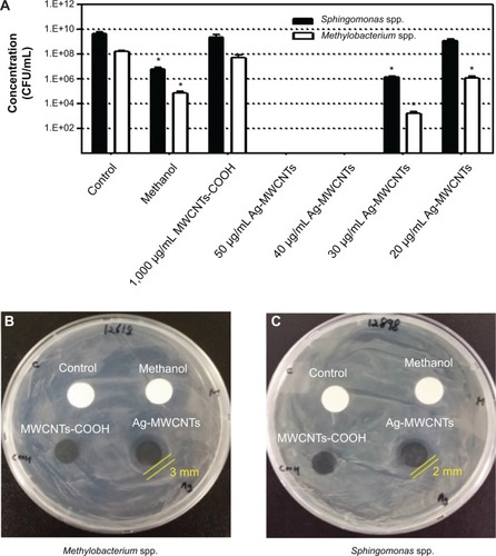
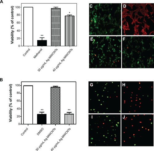
Figure 4 Cell viability assays for 30 and 40 μg/mL Ag-MWCNTs.
Notes: (A and B) AML 12 and human PBMCs were cultured with Ag-MWCNTs for 8 hours and their viabilities analyzed. Viability: *P<0.05, **P<0.01, one-tailed Mann–Whitney U-test. Data are representative of three experiments. (C–F) Photomicrographs of AML 12 cells, fluorescently labeled for dead (red) and live (green) cells. (C) Control; (D) methanol; (E) 30 μg/mL Ag-MWCNTs; (F) 40 μg/mL Ag-MWCNTs. (G–J) Photomicrographs of PBMCs, fluorescently labeled for dead (red) and live (green) cells. (G) Control; (H) DMSO; (I) 30 μg/mL Ag-MWCNTs; (J) 40 μg/mL Ag-MWCNTs.
Abbreviations: DMSO, dimethyl sulfoxide; MWCNT, multi-walled carbon nanotube; PBMCs, peripheral blood mononuclear cells.