Figures & data
Figure 1 The first USA Extra Vehicular Activity (EVA) June 3, 1965, Edward White, (Astronauts Edward White and James McDivitt, Gemini 4, NASA). During the 21 minute long spacewalk, Ed White was tethered to the Gemini spacecraft by a 25 foot “umbilical cord” through which oxygen was supplied. The tethering cord also carried communications and biomedical instrumentation. Note the fine mesh surrounding the cord enhancing flexibility and strength and overall the striking similarity of the supply cord to the human umbilical cord. Interestingly, similar to the umbilical cord the NASA system is tethered to the mid-torso (left mid abdomen) of the astronaut.

Figure 2 Absent umbilical cord in a (twin reverse arterial perfusion TRAP) twin embedded within the placenta. Note the absence of an umbilical cord to this fetus.
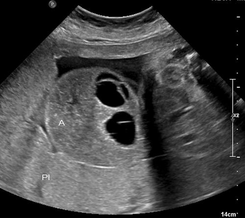
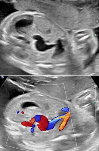
Figure 3 Umbilical cord of thoraco-omphalopagus twins at 23 weeks’ gestation. Upper panel: Axial image of the umbilical cord of thoraco-omphalopagus twins at 23 weeks’ gestation. Note the single umbilical vein and four (4) umbilical arteries (vessels are depicted en face). Lower panel: Doppler energy imaging of thoraco-omphalopagus twins at 23 weeks’ gestation. This image depicts a sagittal view of the umbilical vein, and adjacent four umbilical arteries. As in upper panel, note the single umbilical vein and four (4) umbilical arteries.
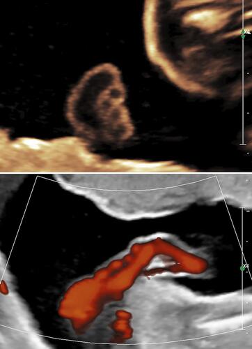
Figure 4 Monochorionic diamniotic twin gestation with reverse twin arterial perfusion (TRAP) sequence at 32 and 1/7 weeks’ gestation. Upper panel: Note furcate, marginal insertion of umbilical cord of the pump (normal twin). Lower panel: The umbilical cord of the pump twin feeds directly to the umbilical cord of the acardiac twin’s placenta. The short and hypocoiled umbilical cord of the acardiac twin (image) contains two vessels and does not communicate with the placenta.
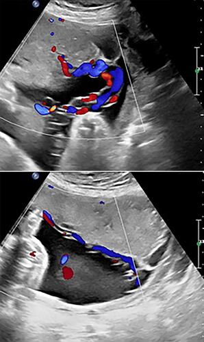
Figure 5 Post delivery image of the placenta and respective umbilical cords of a monochorionic diamniotic twin gestation with reverse twin arterial perfusion (TRAP) sequence delivered spontaneously at 38 weeks’ gestation. Note marginal furcate insertion of the umbilical cord () of the pump (normal twin) on the right. The umbilical cord of the pump twin feeds directly to the umbilical cord of the acardiac twin’s placenta. The umbilical cord of the acardiac twin contains two vessels and does not communicate with the placenta ().
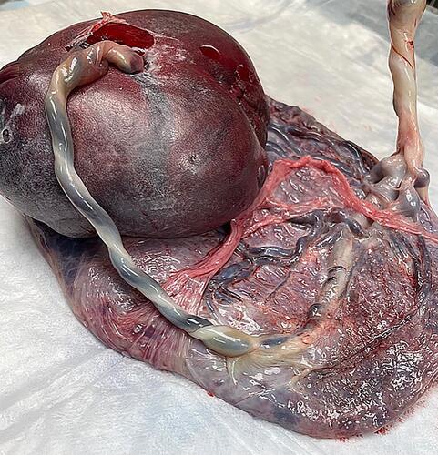
Figure 6 Upper panel: Real-time sonography depicting echogenic mass (*)within the umbilical vein, considered consistent with umbilical vein thrombosis. Lower panel: Color Doppler sonography depicting echogenic mass within the umbilical vein, considered consistent with umbilical vein thrombosis. Black arrows point to the narrow venous inflow and outflow areas, respectively.
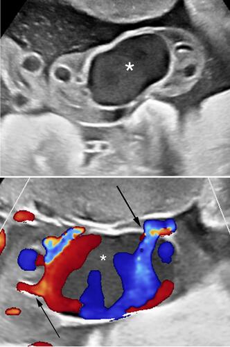
Figure 7 Upper panel: Three-dimensional sonography of the umbilical vein thrombosis. Lower panel: Umbilical vein thrombosis immediately following emergency Cesarean for prolonged fetal bradycardia at 37 weeks’ gestation.
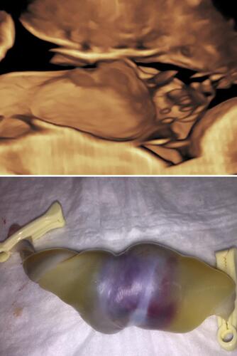
Figure 8 Fetal intra-abdominal varix at 24 and 5/7 weeks’ gestation. Upper panel: Real-time ultrasound depicting the intra-abdominal aneurysmal dilatation of the umbilical vein. Lower panel: Color Doppler imaging confirming the venous vascular nature of this lesion.