Figures & data
Table 1 Recent Cases of Spontaneous Pneumorachis and Management Reported in Literature
Box 1 List of Laboratory Investigations
Figure 1 Chest X-ray PA view of the patient on the day of admission shows a “continuous diaphragm sign” characterised by a mediastinal gas outlining the superior surface of the diaphragm and separating it from the heart (black arrowheads) and a “Naclerio’s V sign” in which mediastinal gas outlines the lateral margin of the descending aorta and extends laterally over the left hemidiaphragm (red arrowheads).
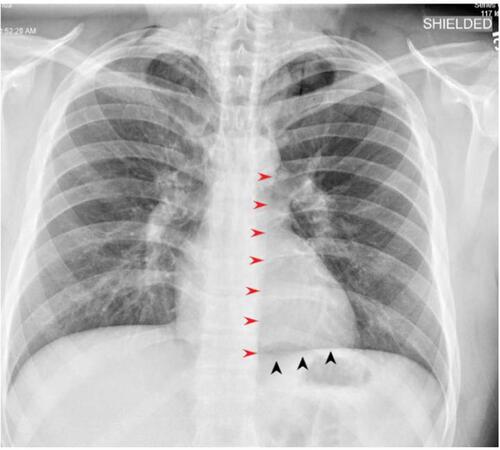
Figure 2 Chest X-ray (lateral view) demonstrating lucency (Yellow arrows) overlying the heart signifying pneumopericardium.
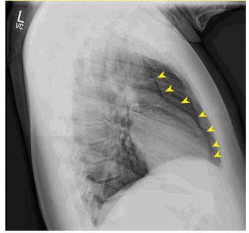
Figure 3 CT imaging demonstrating dissection of fascial planes in neck and invasion of trapped air into the spinal canal (yellow arrows) via intervertebral foramen.
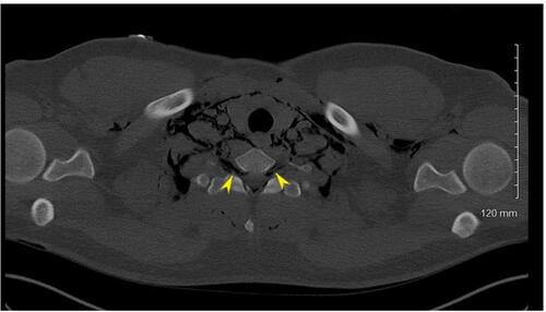
Figure 4 Pneumorachis demonstrated in axial (black arrow) (A) and sagittal sections (B) of thoracic CT imaging (yellow arrow).
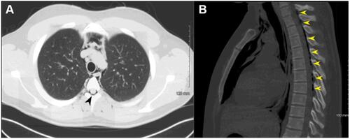
Figure 5 STROBE diagram depicting the selection process stepwise during the literature search for articles on nonspontaneous pneumorachis.
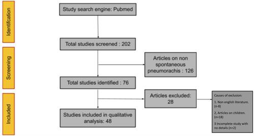
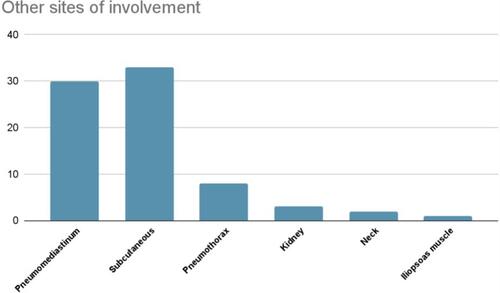
Figure 6 Various sites of air leaks in addition to pneumorachis in our study cohort.