Figures & data
Table 1 Clinical and Functional Characteristic of Type-2 High Severe Asthma Patients with and without Bronchiectasis
Figure 1 Study flow chart.
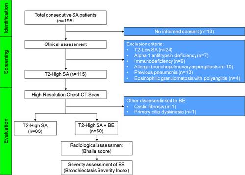
Figure 2 Differences between Type-2 High Severe Asthma patients with and without Bronchiectasis with regard to chronic rhinosinusitis (A), chronic rhinosinusitis with nasal polyps (B), chronic sputum production (C), chronic oral corticosteroids intake (D) and asthma exacerbations in the previous year (E). Receiver operating characteristics (ROC) of the optimal regression model of variables (presence of chronic rhinosinusitis, chronic mucus hypersecretion and daily OCS intake) strongly associated with comorbid BE in T2-SA (F).
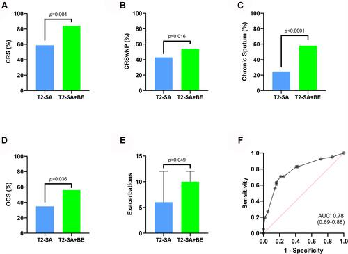
Table 2 Radiological and Microbiological Features in Patients with T2-SA+BE
Figure 3 Bronchiectasis lobes involvement (A) and distribution pattern (B).
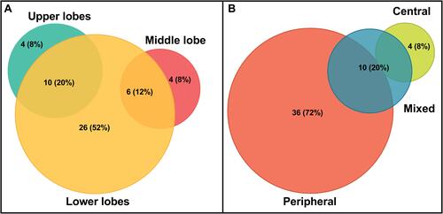
Figure 4 Distribution of patients in relation to Bhalla score items. Data as presented as percentage (%). 1. Severity of bronchiectasis: 0 = None; 1 = luminal diameter slightly larger than the adjacent vessel; 2 = bronchial diameter between 2 and 3 times the diameter of the adjacent vessel; 3 = bronchus is more than 3 times the diameter of the adjacent vessel. 2. Peribronchial thickening: 0 = None; 1 = wall thickness is similar to that of the surrounding vessel; 2 = wall thickness greater, but less than twice, the diameter of the adjacent vessel; 3 = more than twice the thickness of the adjacent vessel. 3. Extent of bronchiectasis: 0 = None; 1 = 1–5 segments; 2 = 6–9; 3 = >9. 4. Mucus plugs: 0 = None; 1 = 1–5 segments; 2 = 6–9; 3 = >9. 5. Sacculation/abscesses: 0 = None; 1 = 1–5 segments; 2 = 6–9; 3 = >9. 6. Generations of bronchial division involved: 0 = None; 1 = >4a generations; 2 = >5a; 3 = >6a. 7. Bullae: 0 = None; 1 = unilateral; 2 = bilateral; 3 = >4. 8. Emphysema: 0 = None; 1 = 1–5 segments; 2 = >5. 9. Collapse/consolidation: 0 = None; 1 = subsegmental; 2 = segmental/lobar.
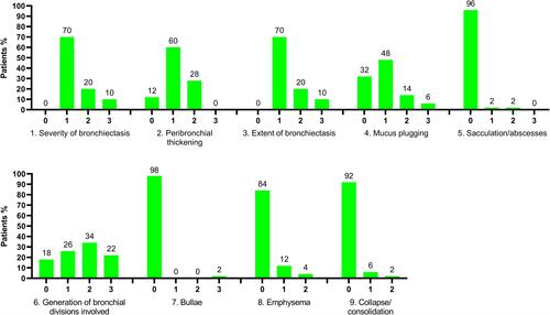
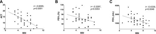
Figure 5 Correlations between BSI, ACT (A), FEV1% (B) and FEV1 mL (C).