Figures & data
Figure 1 The different types of DIC and their clinical presentation. If there is predominance of coagulation pathway activation (denoted as C), in comparison with the fibrinolytic pathways (denoted as F), procoagulant DIC is the result. While the reverse leads to hyperfibrinolytic DIC.
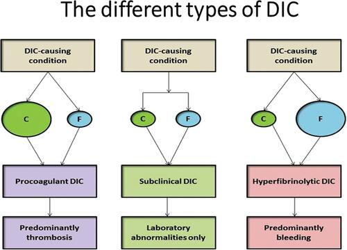
Figure 2 Obstetrical complications associated with DIC in pregnancy.
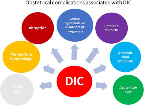
Figure 3 Global distribution of obstetrical complications associated with DIC in pregnancy.
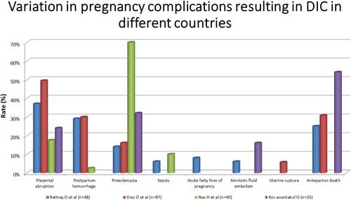
Figure 4 Schematic representation of pathogenic pathways in DIC.
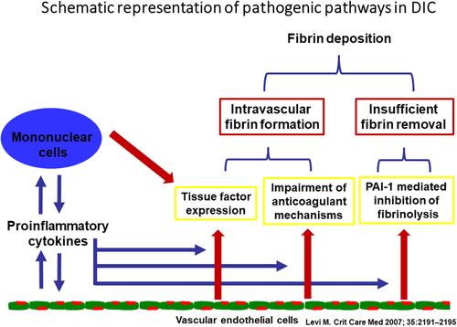
Figure 5 Mechanisms of DIC in sepsis involving endothelial dysfunction and platelet activation.
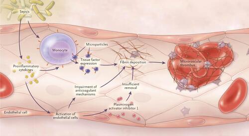
Figure 6 Trophoblast deportation and systemic activation of maternal coagulation cascade. Coagulation system is activated in the placental circulation in the normal pregnant state. This activated coagulation can spill into the peripheral circulation in patients who have obstetrical complications like pre-eclampsia. If coagulation activation in the peripheral circulation becomes uncontrolled, it manifests as DIC.
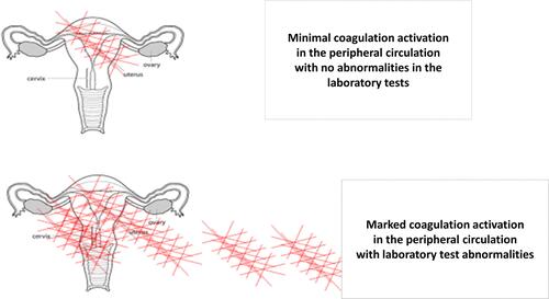
Table 1 Adaptive Changes of the Coagulation System During Pregnancy
Table 2 An Effect of Components of the New DIC Score – Results of Logistic Regression
Figure 7 The changes in the major components of the pregnancy modified DIC score: (A) PT difference: (stands for the difference between the patients PT results and the laboratory control); (B) platelets; (C) fibrinogen, with advancing gestations in women with advancing gestation. (D) ROC curve analysis for the association of the pregnancy specific DIC score with the development of DIC.
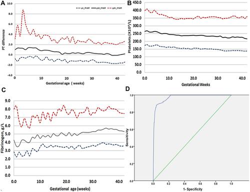
Figure 8 Principles of Diagnosis and management of DIC in pregnancy.
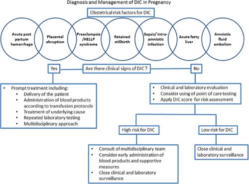
Figure 9 Algorithm for the treatment of obstetrical hemorrhage based on FIBTEM and fibrinogen concentrations.
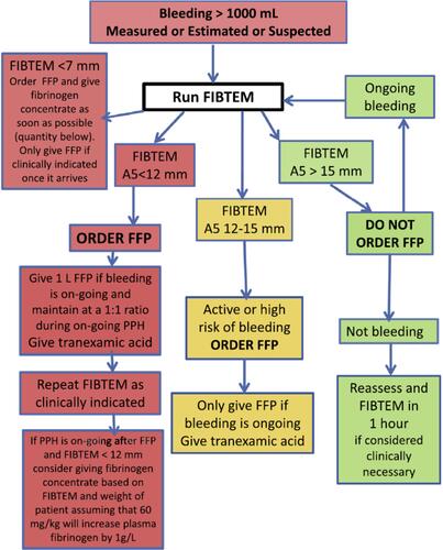
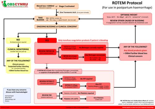
Figure 10 The ROTEM protocol of the OBS Cymru (the Obstetric Bleeding Strategy for Wales) initiative.