Figures & data
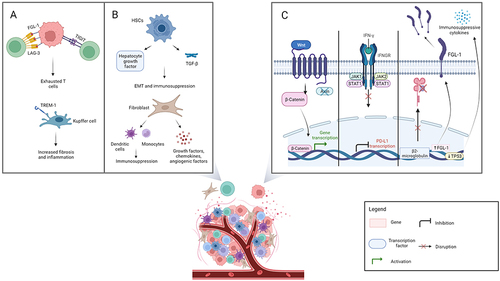
Figure 1 Selected mechanisms of resistance: extrinsic and intrinsic mechanisms are shown in panel A and B, and panel C respectively. Panel A represents the processes involved in T cell exhaustion, which are mediated by lymphocyte activation gene-3 and fibrinogen-like protein 1 (FGL-1) interaction, T cell immunoglobulin and immunoreceptor tyrosine-based inhibitory motif domain expression, and the development of inflammation led by Kupffer cells. Panel B shows the role of activated hepatic stellate cells and fibroblasts in immunosuppression. Panel C depicts intrinsic mechanisms of resistance (from left to right): the Wnt/beta-catenin pathway, interferon γ signaling with Janus kinase / signal transducer and activator of transcription pathway activation, and FGL-1 over-expression with beta-2-microglobulin dysfunction and p53 tumor suppressor gene-mediated immunosuppression. Created with BioRender.com.