Figures & data
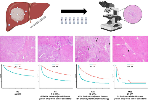
Figure 1 Diagnostic criteria of the MVI grading system. (A) M0; (B). M1; (C) M2a; (D). M2b; (E) Hep Par-1; (F). Arginase-1; (G) Glypican-3; (H) CD34; Arrow: MVI; Magnification of all images: 40×.
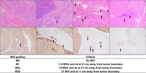
Table 1 Baseline Characteristics of All Patients
Figure 2 Survival analysis of HCC patients based on the MVI grading system. (A) RFS; (B) OS; (C) Early RFS; (D) Late RFS.
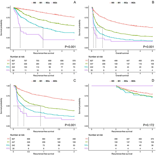
Figure 3 Survival analysis of HCC patients with different amounts of proximal MVI in the M2b group. (A) RFS; (B). OS.
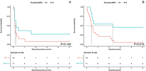
Figure 4 Multivariate Cox regression analysis of prognosis and forest plots. (A) RFS; (B); OS; (C) early RFS; (D) late RFS.
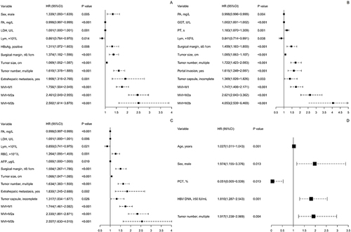
Table 2 Multivariate Logistic Analysis for the Related Factors of MVI
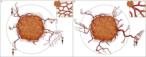
Figure 5 Two kinds of occurrence patterns of distal MVI. (A) Pattern I: the push-out mode. An evolving tumor gradually invades into multifocal vascular wall and spreads from close to distant parts from the tumor boundary. (B) Pattern II: the rush-out mode. A cluster of highly metastatic tumor cells firstly break into a blood vessel within or near the tumor, thus directly causing a distal dissemination of MVI from a sporadic proximal MVI. Arrow: distal MVI; Dotted line: 1 cm from tumor boundary.