
Figures & data
Figure 1 Flowchart of the study. HCC, hepatocellular carcinoma; LR-M, LI-RADS M.
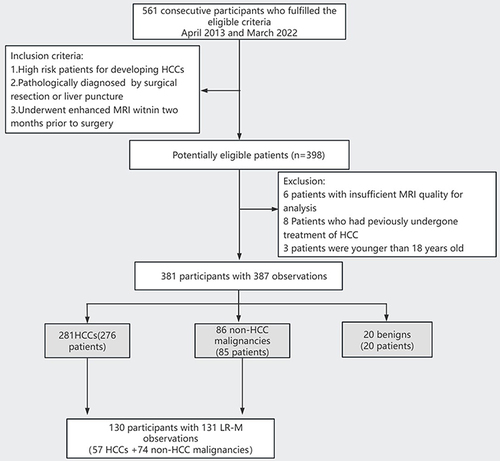
Table 1 Demographic and Pathological Characteristics of 381 Patients with 387 Hepatic Tumors
Figure 2 Rim hyperenhancement patterns on AP image and the modified measurement criterion of rim APHE. (a) Rim hyperenhancement patterns on AP image, thick-rim(left) and thin-rim(right). (b) the modified measurement criterion of rim APHE. Hepatic observations with rim APHE (spatially defined subtype of APHE in which arterial phase enhancement is most pronounced in the periphery) were divided into two categories, thick-rim (30–70%) and thin-rim APHE (< 30%). The thickness measurement of the rim APHE compared with the tumor radius is T/R=30%-70%, classified as thick-rim APHE (left). The thickness measurement of the rim APHE compared with the tumor radius is T/R<30%, classified as thin-rim APHE(right).
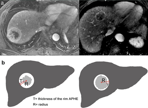
Table 2 Liver Imaging-Reporting and Data System (LI-RADS v2018) Categorization of HCC and Non-HCC Malignancies Using Contrast-Enhanced MRI
Table 3 MRI Features of Malignant Observations According to MRI Modality (ECA-MRI Vs HBA-MRI)
Table 4 Comparison of LI-RADS Characteristic of the Study Population Within LR-M Category
Figure 3 Hepatocellular carcinoma in a 74-year-old man with chronic hepatitis B. (a-c) On the axial arterial phase, portal venous phase and delayed phase images after administration of extracellular contrast agent, the segment-5 lesion showed thick-rim arterial phase hyperenhancement (APHE) followed by non-peripheral washout. (d) The mass showed a non-targetoid appearance on b-800 diffusion weighted image. The tumor was initially categorized as LR-M by observers. The tumor was re-categorized as modified LR-5 according to modified LI-RADS.
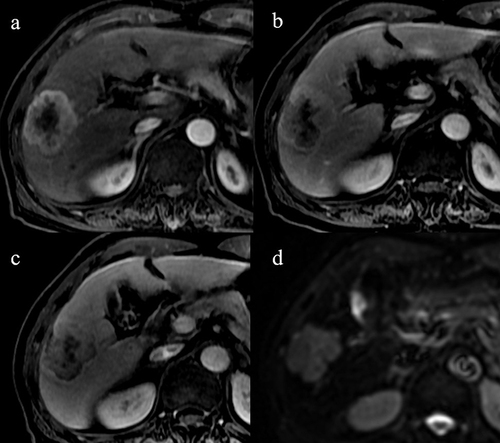
Figure 4 Intrahepatic cholangiocarcinoma in two patients, a 48-year-old man with chronic hepatitis B. On the axial arterial phase image (a), the liver mass on segment 5 showed thin-rim arterial phase hyperenhancement (APHE). On portal venous phase and delayed phase images (b and c) after administration of extracellular contrast agent, the tumor showed delayed central enhancement. On b-800 DWI image (d), the mass showed a targetoid appearance. The tumor was initially categorized as LR-M by observers. The tumor was re-categorized as modified LR-M according to modified LI-RADS.
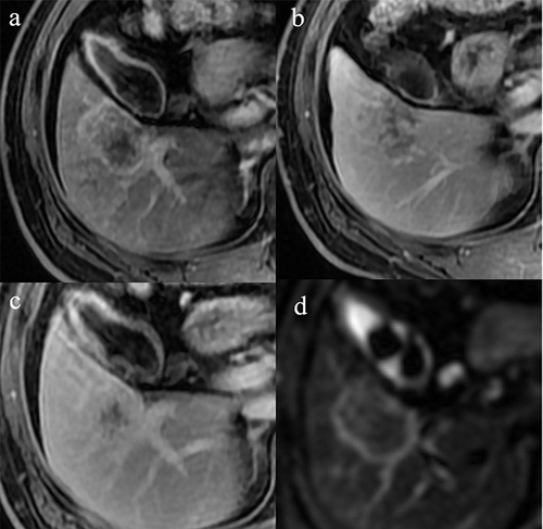
Figure 5 Performance of thin-rim APHE, targetoid DWI and peripheral washout to distinguish non-HCC malignancy from HCC in targetoid lesions.
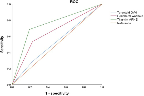
Table 5 Diagnostic Performance of LI-RADS Categories for the Diagnosis of Hepatic Malignancies Using Enhanced MRI