Figures & data
Figure 1 Flowchart of the study population.
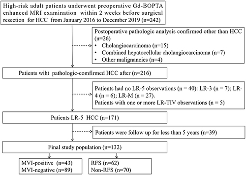
Table 1 Detailed Sequences and Parameters of Gd-BOPTA Enhanced MRI
Table 2 Clinicopathologic Characteristics of the Study Population
Table 3 Univariate and Multivariate Logistic Analyses for MVI with Patients of LR-5 HCC
Table 4 Univariate and Multivariate Cox Analyses for Recurrence with Patients of LR-5 HCC
Figure 2 Kaplan–Meier curves of imaging features for RFS in LR-5 HCCs. (A) Mosaic architecture (absent) and Mosaic architecture (present) (Log rank test, p =0.034). (B) Corona enhancement (absent) and Corona enhancement (present) (Log rank test, p <0.0001). (C) Peritumoral hypointensity on HBP (absent) and Peritumoral hypointensity on HBP (present) (Log rank test, p <0.0001).
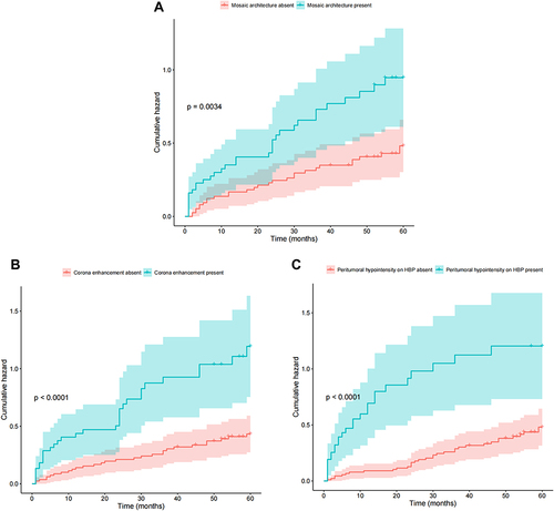
Table 5 Diagnostic Performance for Predicting MVI and Recurrence
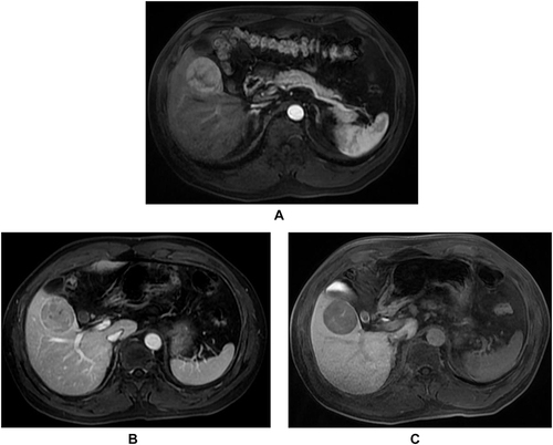
Figure 3 Images of a 54-year-old man with chronic hepatitis B infection, categorized as LR-5 HCC with MVI and recurrence. (A) Heterogeneous mass with mosaic architecture on T2WI; (B) Non-rim arterial phase hyperenhancement (APHE) on the arterial phase, with corona enhancement and internal arteries (white arrow). (C) Peritumoral hypointensity (white arrow) in the hepatobiliary phase.

Figure 4 MR images of a 48-year-old man with chronic hepatitis B infection, categorized as LR-5 HCC without MVI and non-recurrence. (A) Non-rim arterial phase hyperenhancement (APHE) on the arterial phase. (B) Non-peripheral washout on the portal venous phase. (C) The lesion of hypointense on the HBP image.