Figures & data
Table 1 The Primer Sequences for qRT-PCR
Figure 1 KCNQ1OT1 was up-regulated and miR-30e-3p was down-regulated in LPS-induced HMC3 cells. HMC3 cells were treated with different concentrations of LPS (0, 0.1, 0.2, 0.5 and 1 μg/mL) for 24 h, or treated with 1 μg/mL of LPS for different times (0, 1, 6, 12 and 24 h). RT-qPCR was used to detect the expression levels of KCNQ1OT1 (A and B) and miR-30e-3p (C and D) in HMC3 cells. All experiments were performed in triplicate. *P<0.05, **P<0.01, and ***P<0.001.
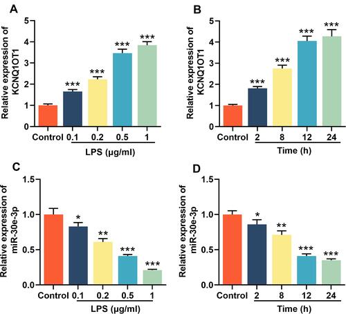
Figure 2 Knocking down KCNQ1OT1 inhibited LPS-induced neuroinflammation and neuronal apoptosis in HMC3 cells. HMC3 cells were transfected with pc-NC, pc-KCNQ1OT1, si-NC or si-KCNQ1OT1, respectively, and HMC3 cells were treated with 1 μg/mL LPS for 24 h after transfection. (A) RT-qPCR was used to detect KCNQ1OT1 expression. (B) RT-qPCR was used to detect the expression level of TNF-α, IL-1β and IL-6 mRNA. (C) ELISA was used to detect TNF-α, IL-1β and IL-6 in the cell culture supernatant of HMC3 cells. (D and E) RT-qPCR was used to detect the expression level of CD86 and CD206. (F) MTT assay was used to detect the viability of HMC3 cells. (G) The neuronal injury was detected using LDH cytotoxicity detection kit. (H and I) The levels of NO and ROS were detected. All experiments were performed in triplicate. *P<0.05, **P<0.01, and ***P<0.001.
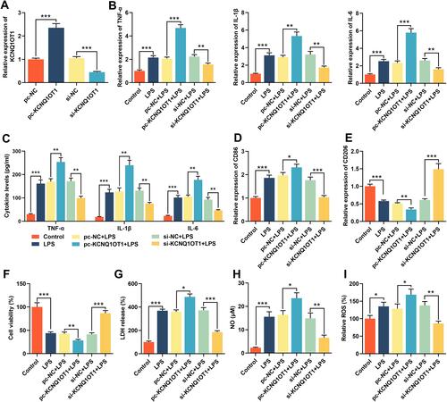
Figure 3 miR-30e-3p was the target of KCNQ1OT1. (A) Lncbase database was used to predict the binding site for miR-30e-3p in KCNQ1OT1 sequence. (B) HMC3 cells were co-transfected with miR-30e-3p mimics or control mimics and luciferase reporter vector KCNQ1OT1-WT or KCNQ1OT1-MUT, and luciferase activity of the cells in different group was detected after 48 h. (C) RT-qPCR was used to detect the expression of miR-30e-3p in HMC3 cells transfected with pc-KCNQ1OT1 or si-KCNQ1OT1. All experiments were performed in triplicate. **P<0.01 and *** P <0.001.
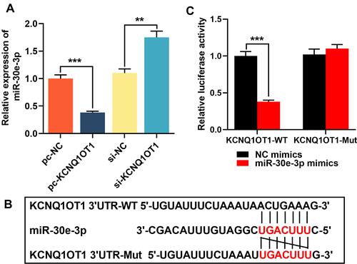
Figure 4 KCNQ1OT1 reversed the inhibitory effect of miR-30e-3p on LPS-induced neuroinflammation and neuronal apoptosis in HMC3 cells. MiR-NC, miR-30e-3p mimics, miR-30e-3p mimics + pc-NC or miR-30e-3p mimics + pc-KCNQ1OT1 were transfected into HMC3 cells, respectively. (A) The expression level of miR-30e-3p was detected by RT-qPCR. (B) RT-qPCR was used to detect the expression level of TNF-α, IL-1β and IL-6 mRNA. (C) ELISA was used to detect TNF-α, IL-1β and IL-6 in the cell culture supernatant of HMC3 cells. (D and E) RT-qPCR was used to detect the expression level of CD86 and CD206. (F) MTT assay was used to detect the viability of HMC3 cells. (G) The neuronal injury was detected using LDH cytotoxicity detection kit. (H and I) The levels of NO and ROS were detected. All experiments were performed in triplicate. *P<0.05, **P<0.01 and ***P<0.001.
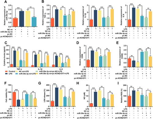
Figure 5 KCNQ1OT1 indirectly promoted NLRP3 expression by inhibiting miR-30e-3p expression. (A) miRanda database was used to predict the binding site between miR-30e-3p and NLRP3 3ʹUTR sequence. (B) HMC3 cells were co-transfected with miR-30e-3p mimics or control mimics and luciferase reporter NLRP3-WT or NLRP3-MUT, and the luciferase activity of the cells in different groups was detected after 48 h. (C and D) RT-qPCR and Western blot were employed to detect the expression level of NLRP3 mRNA and protein in HMC3 cells after KCNQ1OT1 and miR-30e-3p were selectively regulated. All experiments were performed in triplicate. *P<0.05, **P<0.01 and ***P<0.001.
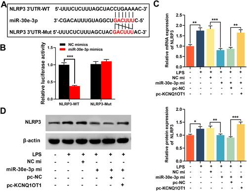
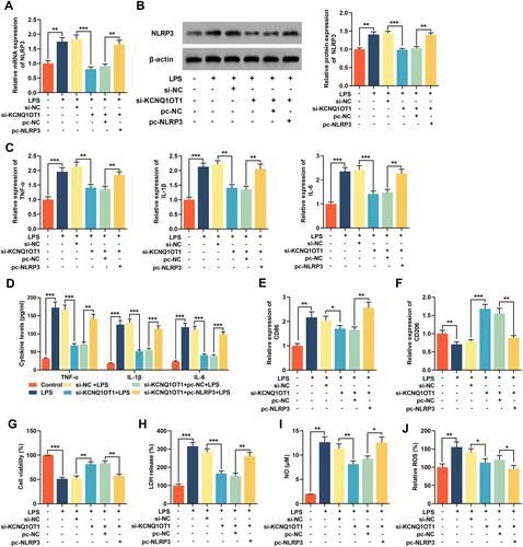
Figure 6 Overexpression of NLRP3 reversed the inhibitory effect of knocking down KCNQ1OT1 on LPS-induced neuroinflammation and neuronal apoptosis in HMC3 cells. The HMC3 cells were transfected with si-NC, si-KCNQ1OT1, si-KCNQ1OT1+pc-NC or si-KCNQ1OT1+pc-NLRP3, respectively. After 48 h of transfection, HMC3 cells were treated with 1 μg/mL LPS for 24 h. (A and B) The expression level of NLRP 3 was detected using RT-qPCR and Western blot. (C) RT-qPCR was used to detect the expression level of TNF-α, IL-1β and IL-6 mRNA. (D) ELISA was used to detect TNF-α, IL-1β and IL-6 in cell culture supernatant of HMC3 cells. (E and F) RT-qPCR was used to detect the expression level of CD86 and CD206. (G) MTT assay was used to detect the viability of HMC3 cells. (H) The neuronal injury was detected using LDH cytotoxicity detection kit. (I and J) The levels of NO and ROS were detected. All experiments were performed in triplicate. *P<0.05, **P<0.01 and ***P<0.001.