Figures & data
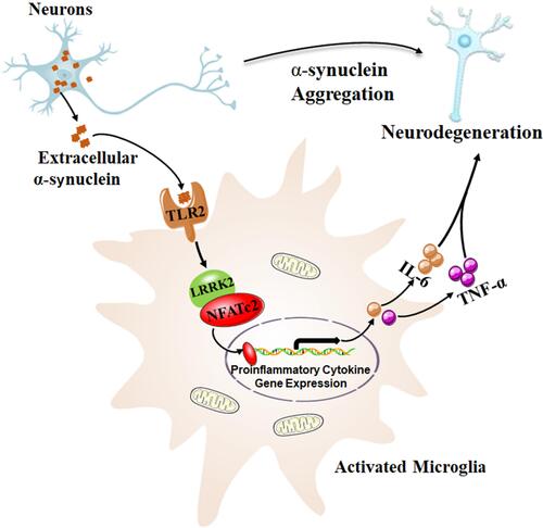
Figure 1 Neuron-released α-synuclein activated microglia through TLR2, and subsequently activated LRRK2 kinase. NFATc2, as a kinase substrate of LRRK2, was directly phosphorylated, which accelerated nuclear translocation of NFATc2 where cytokine/chemokine gene expression such as IL-6 and TNF-α, were upregulated by NFATc2 transcriptional activity. This resulted in a neurotoxic inflammatory environment, which in return aggravated the neuronal degeneration in a mouse model of PD. Data from Kim et al.Citation15