Figures & data
Table 1 Patients’ Clinical Data
Table 2 Classification of SPINs Based on Key CT Features
Table 3 Classification of Lung Cancers Based on Key CT Features
Figure 1 Solid SPINs with smooth margin (type Ia) (A), coarse margin (type Ib) (B), sparse and long spiculations (type Ic) (C), and sparse and short spiculations (type Ic) (D). Pathologically, they have similar manifestations including significant fibrous tissue proliferation, hyaline change and few chronic inflammatory cells infiltration (E–H).
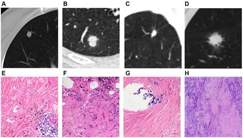
Figure 2 Solid SPINs with blurred margin (A) and peripheral patch (C) (type II). Pathologically, they have similar manifestations including more acute and chronic inflammatory cells infiltration and fibrous tissue proliferation (B and D).
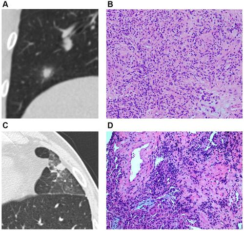
Figure 3 Solid SPIN with heterogeneous density (type III) and spiculations (A) on lung window. Pathologically, it consists of multiple components including fibrous tissue (B), hyaline change (C), calcification (D), and few inflammatory cells (B and C).
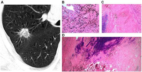
Figure 4 Solid SPIN with polygonal shape (type IV) (A). It has homogeneous density and smooth margin. Pathologically, it consists of fibrous tissue proliferation, hyaline change and a small amount of inflammatory cells infiltration (B).
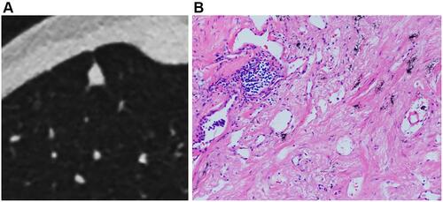
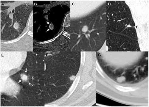
Figure 5 Solid cancerous nodules with smooth and lobulated margin (type Ia) (A), coarse margin (type Ib) (B), lobulated margin and sparse and long spiculations (type Ic) (C), intensive and short spiculations (type Ic) (D), well-defined peripheral patch locating at one side (arrow) (E) or surrounding lesion (arrows) (F) (type II), and heterogeneous density (type III) (G and H).
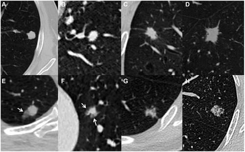
Table 4 The Pathological Findings of SPINs
Figure 6 Tuberculous nodules with smooth margin (A) and curved calcification (arrows) (B), coarse margin (C), sparse and long spiculations and pleural indentation (arrow) (D), and ill-defined peripheral patch (arrows) (E). A cryptococcal nodule abutting pleura with a wide base (F), and an oval aspergillus nodule with clear and smooth margin (G).