Figures & data
Figure 1 Standard multicellular epidermal culture model in which the cells are grown on a polycarbonate filter (Filter I) which is placed on a sponge (Sponge I). The sponge is kept in a culture medium in a Petri dish. Capillary diffusion of the medium transport liquid medium up to the filter which is used to nourish the cells (A). If the distance between the culture medium and the cells is extended by adding one extra sponge (sponge II), capillary osmosis decreases and the liquid supply to the cells diminishes (B). Exposing the multicellular skin cell surface with a hypertonic liquid (e.g. glycerol), should improve cell survival due to the higher osmotic forces exerted by the hypertonic liquid.

Table 1 Percentage Increase (+) or Decrease (-) in Cell Survival of Epidermal Cells in 2-Sponges Cellular Dehydration Model When Exposed to 10% Glycerol Containing 0.10% Glycerol Binding Polymer(s) vs. 10% Glycerol Alone. The Cell Membranes Were Exposed to Mechanical Pressure at 6 h, 24 h, and 48 h by Stirring in Culture Medium. Higher Cell Survival Indicates Higher Film Resistance to Mechanical Pressure and Better Filmogenicity. The Polymeric Association Incorporated in Covispray Were Called S1 cyanidins and Was Selected Among the Key Polymers (AvL,CsL, ClR, UdP, TpF, PgR, HhP, VmaFr) Derived from Tannin-Rich Whole Plant Extracts. Gly, Glycerol; HpP, Hydroxy Propyl Cellulose; Sg, Solagum (an Association of Acacia and Xanthan Gum) Were Used as Absorbent Excipients
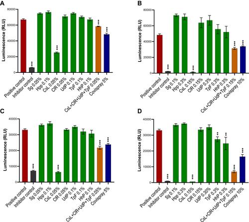
Figure 2 SARS-CoV2 spike S1 or RBD proteins were incubated with test products in low (A and C) or high (B and D) concentrations and were exposed onto ACE2 receptor protein coated ELISA assay microwells. Positive controls had no interactions with virus proteins while negative inhibitor controls were designed to bind with virus proteins. Polymeric ingredients or their associations were tested either in concentrations between 0.05–0.10% (A and C) or between 0.10–0.30% (B and D) while the finished product formulation was tested at 5% or 10% concentration. Results are presented as mean (n=4) viral protein inhibitions against S1 protein (A and B) or RBD protein (C and D). Luminescence was analyses using one-way ANOVA followed by Sidak’s post hoc test. Confidence intervalles 95%. **p<0.01 ***p<0.001.

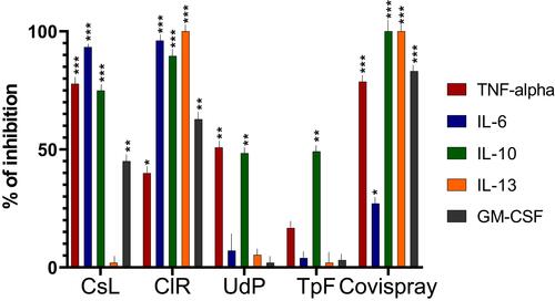
Figure 3 The key Covid-19 disease specific pro-inflammatory cytokines (TNF-α, IL-6, IL-10, IL-13 and GM-CSF) were incubated for 5 min with four polymers (CsL, CIR, UdP, and TpF) at a concentration of 0.10% and with the finished product at a concentration of 5.0%. Percentage inhibition of cytokine activity was evaluated with ELISA sandwich assays (n = 18 per test concentration). Results were statistically analyzed using one-way ANOVA followed by Dunnett’s post hoc tests. Confidence intervals 95%. *p<0.05 **p<0.01 ***p<0.001. Results show that CsL associated with CIR polymers can block between 60–90% of all Covid-19 specific inflammatory proteins. The best cytokine and viral protein binding polymers were then associated to conceive Covispray.

