Figures & data
Figure 1 Ac-SDKP reduces formation of TRAP-positive macrophages. TRAP staining, Bar=2 mm and 500 μm, Data are presented as the mean ± SD. n = 10 per group.
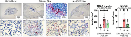
Figure 2 Ac-SDKP reduces activation of RANKL signaling in rats exposed to silica. (A) Co-expression of RANK/CD68 in rats exposed to silica, Bar=50 μm; (B) Co-expression of RANKL/CD68 in rats exposed to silica, Bar=50 μm; (C) Levels of RANKL, RANK, OC-STAMP, AP-1, NFATc1, and NF-κB in rats lungs measured by Western blotting. Data are presented as the mean ± SD. n = 3 per group.
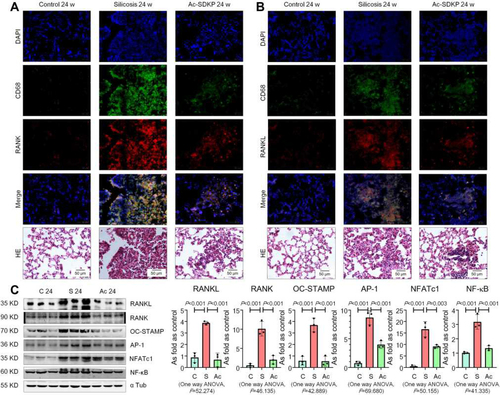
Figure 3 Ac-SDKP reduces lung inflammation and activation of TLR4 signaling in rats exposed to silica. (A) Co-expression of TLR4/CD68 in rats exposed to silica, Bar=50 μm; (B) Co-expression of TNF-α/CD68 in rats exposed to silica, Bar=50 μm; (C) Levels of TLR4. MyD88, p-IκBα, and TNF-α in rats lungs measured by Western blotting. Data are presented as the mean ± SD. n = 3 per group. (D) Levels of TNF-α and RANKL in serum of rats measured by ELISA. Data are presented as the mean ± SD. n = 10 per group.
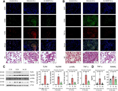
Figure 4 Ac-SDKP inhibits MMP-12 and stabilizes elastin in silicotic rats. (A) Elastin staining, Bar=200 μm, Data are presented as the mean ± SD. n = 8 per group; (B) Protein expression of MMP-12 in rats lungs measured by Western blotting. Data are presented as the mean ± SD. n = 3 per group.
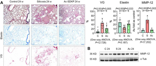
Figure 5 Ac-SDKP inhibits TLR4 and RANKL signaling in NR8383 cells treated with SiO2. (A) TRAP staining, Bar= 50 μm; (B) IF staining of TLR4, Bar=100 μm; (C) Protein expression of RANKL, RANK, OC-STAMP, AP-1, NFATc1, TRAF6, NF-κB, TLR4, MyD88, p-IκBα, TNF-α, and MMP-12 in NR8383 cells treated with SiO2 and Ac-SDKP or not. Data are presented as the mean ± SD. n = 3 per group.
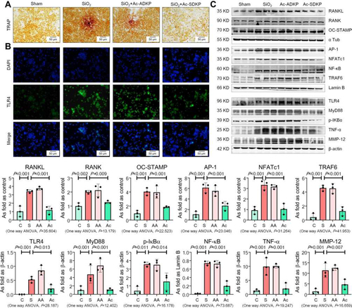
Figure 6 Ac-SDKP reduces TRAP-positive cells and RANKL signaling in RAW 264.7 cells treated with SiO2. (A) TRAP staining, Bar= 500 μm; (B) Levels of OC-STAMP, AP-1, NFATc1, and NF-κB in RAW 264.7 cells treated with 50, 100, and 200 µg/mL silica measured by Western blotting. *Compared with control group, P<0.05. Data are presented as the mean ± SD. n = 3 per group. (C) Protein expression of OC-STAMP, AP-1, NFATc1, and NF-κB in RAW 264.7 cells treated with SiO2 and Ac-SDKP or not. Data are presented as the mean ± SD. n = 3 per group.
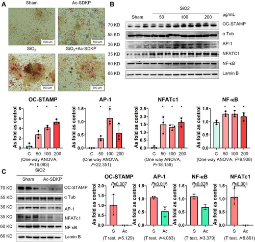
Figure 7 Ac-SDKP reduces TLR4 signaling and MMP-12 in silica-treated macrophages. Levels of TLR4, MyD88, p-IκBα, TNF-α, and MMP-12 in RAW 264.7 cells measured by Western blotting. Data are presented as the mean ± SD. n = 3 per group.
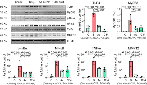
Figure 8 Ac-SDKP inhibits the decrease in BMD in the femur. (A) Bone microstructure in the femur; (B) Bone microstructure in the tibia.
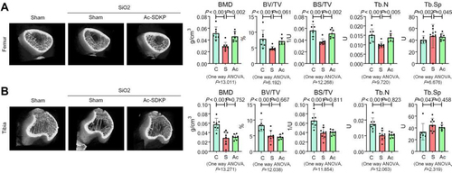
Figure 9 Ac-SDKP inhibits osteoclast differentiation in silicotic rats. (A) TRAP staining and ALP staining in the femur, Bar= 500 μm and 100 μm; (B) TRAP staining and ALP staining in the tibia, Bar= 500 μm and 100 μm.
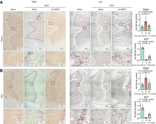
Figure 10 Ac-SDKP inhibits osteoclast differentiation in RAW 264.7 cells treated with RANKL. (A) TRAP staining and Osteo assay, Bar=500 μm. (B) Protein expression of OC-STAMP, AP-1, NFATc1, and NF-κB in RAW 264.7 cells treated with Ac-SDKP in different dose measured by Western blotting. *Compared with control group, P<0.05. Data are presented as the mean ± SD. n = 3 per group. (C) Protein expression of OC-STAMP, AP-1, NFATc1, and NF-κB in RAW 264.7 cells treated with RANKL and Ac-SDKP or not. Data are presented as the mean ± SD. n = 3 per group.
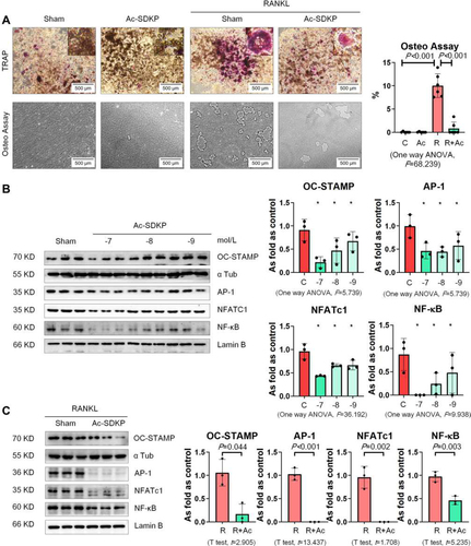
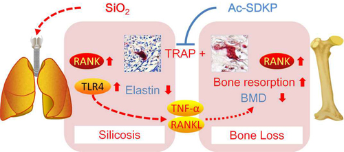
Figure 11 Ac-SDKP attenuates activation of lung macrophages and bone osteoclasts in rats exposed to silica.