Figures & data
Figure 1 Cardiovascular and non-cardiovascular functions of Piezo1: The schematic diagram demonstrating a wide range of Piezo1 functions in different areas of physiology including cardiovascular and non-cardiovascular roles from the activation of Piezo1 by mechanical force branching from left to right arrows, pointing different organ and cell /tissue.
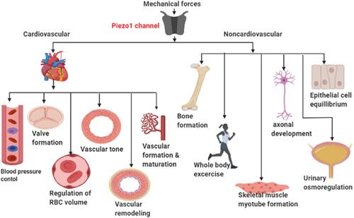
Table 1 Summarizing the Suggested Clinical Significance of Helical Flow
Figure 2 Piezo1 flow induced anti-atherogenic signaling mechanism: schematic diagram, demonstrating laminar flow (anti-atherogenic flow) activates Piezo1 channel, leading to Ca2+ influx and then triggers the generation of eNOS that leads to the expression of KLF2 & KLF4 through MAPK and results in the deactivation of adhesion molecules and AP-1 (red inhibition arrow) as well as downregulations of growth factors VEGF/PDGF, proinflammatory chemokines and cytokines, cell proliferation and migration followed by anti-atherogenic gene expression and athero-protection.
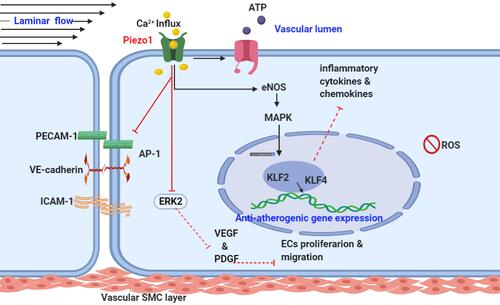
Figure 3 Piezo1 flow induced proatherogenic signaling mechanism: schematic diagram demonstrating disturbed/oscillatory (pro-atherogenic flow) activation of the Piezo1 channel, leading to Ca2+ influx followed by the activation of JNK, NF-KB, p50 and p65, ERK2 pathway. And the atherogenic gene expression, growth factors VEGF/PDGF, proinflammatory chemokines, cytokines. And cellular adhesion molecules, which result in the activation of immune cells, inflammatory cell recruitment, and ECs and SMCs proliferation and migration, leading to endothelial inflammation, increased permeability, cell damage, plaque formation, and atherosclerosis development.
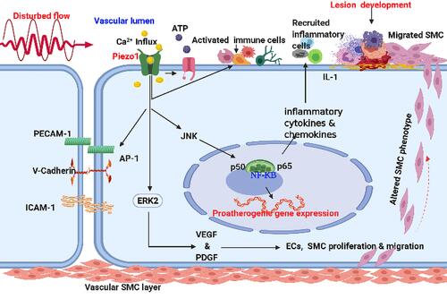
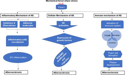
Figure 4 Different mechanosignaling mechanisms of atherosclerosis: schematic diagram demonstrating different signaling thoroughfares and stages of atherosclerotic development from Piezo1 activation by mechanical force, the arrow branching to inflammatory, cellular proliferation and migration, and immune atherosclerosis development.