Figures & data
Figure 1 Induced aGVHD model in NPG mice. (A) Representative clinical manifestations including hunched posture, ruffed fur, damaged skin, and hematochezia in the aGVHD mouse model. (B) Measurement of weight change, clinical score, and survival time at different time points with those two groups. The weight loss rate in aGVHD group is faster than in the control group, the clinical score of aGVHD was higher than that of the control group, the survival time of the control group was also significantly longer than that of the aGVHD group. *p < 0.05 and **p < 0.01. (C) Representative histology of target organs, including the liver, spleen, lung and gut, showed inflammatory cell infiltration and tissue damage in aGVHD group. 400×. *p < 0.05 and **p < 0.01. (D) Representative immunohistochemistry of target organs. Human CD45+ cells were detected by immunohistochemistry in these target organs. 400×. *p < 0.05 and ***p < 0.001. (E) Human CD3+ T cells were not detected by flow cytometry in the control group. Human CD3+ T cells from each target organ were detected by flow cytometry in the aGVHD group.
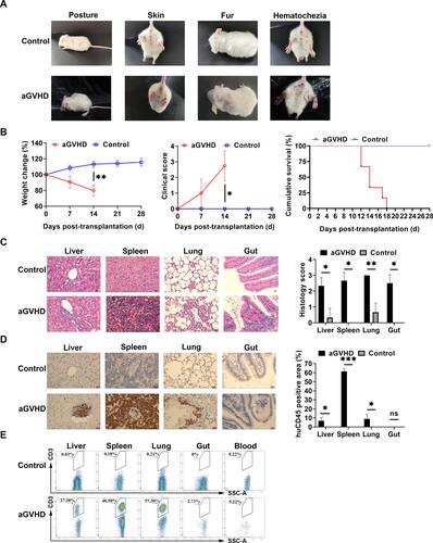
Figure 2 Identification of human amnion mesenchymal stem cells. (A) Characteristics of passage three hAMSCs. (B) Osteogenic differentiation of hAMSCs indicated by Alizarin Red S. 100×. (C) Adipogenic differentiation of hAMSCs indicated by Oil Red O staining. 100×. (D) Surface antigens of hAMSCs were detected by flow cytometry. Cells were positive for CD90, CD105, and CD73 and negative for CD45, CD11b, CD34, and HLA-DR. (E) GFP-labeled hAMSCs and 293T cells were detected by fluorescence microscopy at day 3 following transfection with GFP-pseudovirion. 100×. ***p < 0.001.
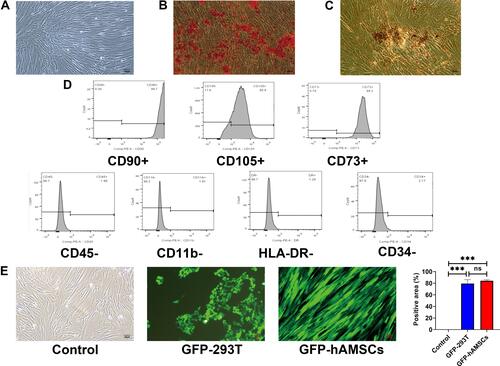
Figure 3 Human amnion mesenchymal stem cells migrated to target organs. GFP-labeled hAMSCs detected by confocal microscopy in target organs at 24 after injection in vivo. At 72 h, GFP fluorescence in the spleen, liver, and gut was weakened while increased in the lung.
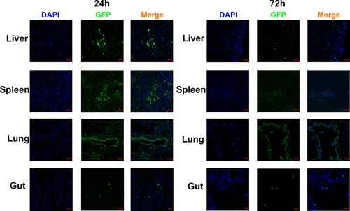
Figure 4 The capacity of hAMSCs inhibited the development of -aGVHD in vitro. (A) Measurement of weight change (aGVHD vs aGVHD+hAMSCs day 7 P=0.043, day 14 P=0.037), clinical score (aGVHD vs aGVHD+hAMSCs day 14 P=0.002), and survival in the different groups (aGVHD vs aGVHD+hAMSCs P<0.001). *p < 0.05 and **p < 0.01. (B) Representative histology of target organs, including the liver, spleen, lung, and gut. Compared with the hAMSCs treatment group, inflammatory cell infiltration and tissue damage in the aGVHD group were more severe. 400×. *p < 0.05 and **p < 0.01. (C) Representative immunohistochemistry of target organs, including the liver, spleen, lung, and gut. Infiltration and tissue damage in the aGVHD group were more severe than that of hAMSCs treatment group and Control group. 400×. **p < 0.01 and ***p < 0.001.
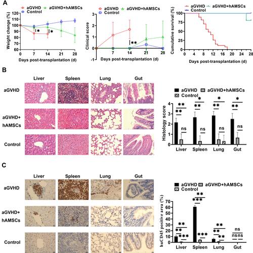
Figure 5 hAMSCs suppressed effector T cell and increased Tregs in target organs. (A) After hAMSCs treatment, the proportion of CD3+CD4+ T cells in liver, spleen and gut was significantly lower than that of aGVHD group. *p < 0.05. (B) After hAMSCs treatment, the proportion of CD3+CD8+ T in liver, spleen, lung and gut was significantly lower than that of aGVHD group. *p < 0.05 and ***p < 0.001. (C) After hAMSCs treatment, the proportion of CD4+CD25+Foxp3+ Tregs in liver, spleen, and gut was significantly increased compared with that of aGVHD group. *p < 0.05.
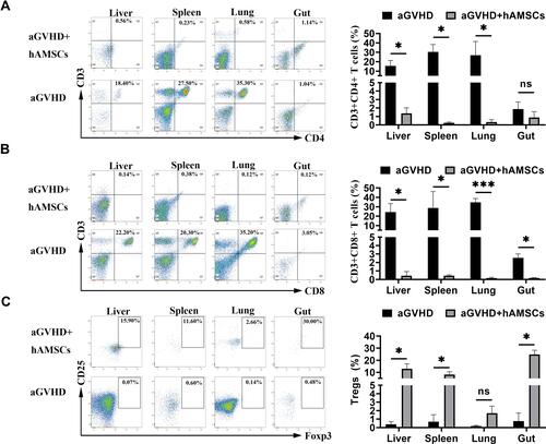
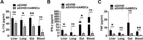
Figure 6 hAMSCs can inhibit the secretion of inflammatory cytokine in target organs. (A) IL-17A level in lung and gut were significantly decreased in aGVHD+hAMSCs group than that in aGVHD group. *p < 0.05 and **p < 0.01. (B) IFN-γ level in liver and gut were significantly decreased in aGVHD+hAMSCs group than that in aGVHD group. *p < 0.05. (C) TNF level in liver and blood were significantly decreased in aGVHD+hAMSCs group than that in aGVHD group. *p < 0.05 and **p < 0.01.