Figures & data
Figure 1 Diagram, showing the overall contents of the manuscript covering the introduction and the contents related to melatonin.
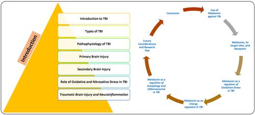
Figure 2 Pathophysiology of traumatic brain injury. A brief illustration of the primary brain injury, which is consisted of stretching, compression, and tearing followed by secondary brain injury which leads to further induce oxidative stress, neuroinflammation, excitotoxicity, apoptotic cell death, blood-brain barrier disruption, dendritic degeneration, synaptic loss, and ultimately progress towards neurodegeneration and cognitive dysfunctions.
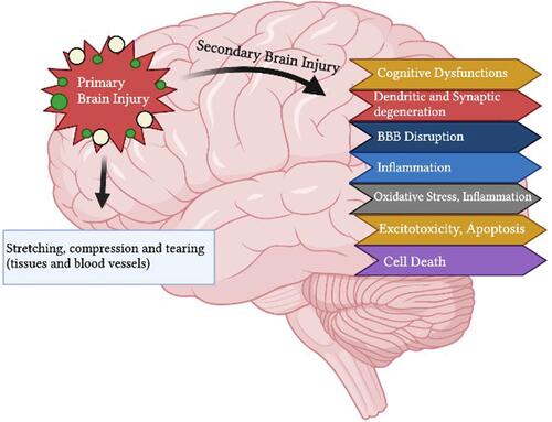
Figure 3 Brain injury-induced oxidative and nitrosative stress and its consequences. Diagrammatic representation, showing that brain injury induces oxidative and nitrosative stress, which causes further deterioration by disrupting the blood-brain barrier, activating the astrocytes and microglial cells, releasing the inflammatory cytokines, infiltration of the lymphocytes, and may cause the accumulation of misfolded proteins and neurodegeneration.
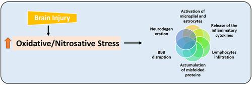
Figure 4 Traumatic brain injury and neuroinflammation. The figure illustrates the activation of microglia (Iba-1 and microglial receptor TLR-4), astrocytes (GFAP), MAP kinases (such as p-JNK and p-38), the release of the inflammatory cytokines (TNF-α and IL-1β), and release of chemokines, which may induce neuroinflammation and neurodegeneration.
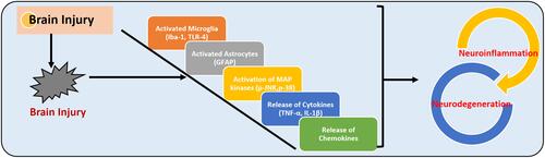
Table 1 Summary of the Drugs Used Against Traumatic Brain Injury-Induced Neurodegeneration, Therapeutic Targets, and Implications
Figure 5 Melatonin is a potent antioxidant in brain injury-induced neurodegeneration.
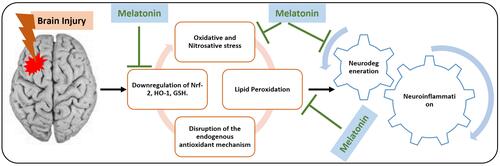
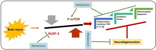
Figure 6 Effects of melatonin against Brain injury-induced activated NLRP-3 inflammasome and autophagic dysfunction. Graphical representation of effects of melatonin against brain injury-induced the activation of NLRP-3 inflammasome and inhibits the phosphorylation of mTOR. As the activated NLRP-3 inflammasome are responsible for the release of inflammatory cytokines, mitochondrial apoptosis, and elevated oxidative stress. Moreover, it has been suggested that melatonin has regulatory effects on the mTOR-mediated autophagy and NLRP-3 inflammasome activation.