Figures & data
Figure 1 Baicalin recovered the bone resorption in the Bone metastases cancer. CT was used to detect BMD in SD rats of the NC group, the Bone metastases group and the Bone metastases + Baicalin group. The changes of BMD were calculated by SPSS software.
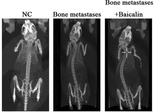
Figure 2 Baicalin inhibited osteoclast activation and promoted the increase of bone trabeculae in the Bone metastases cancer. Trap is a typical marker of osteoclasts, and the results of Trap staining was that the cytoplasm of osteoclasts was wine red and the nucleus was light blue. Trap staining indicated that osteoclasts count was significantly increased in the Bone metastases group and was decreased after Bone metastases + Baicalin. HE staining is one of the commonly used staining methods in paraffin section technology. The nucleus is dyed purple blue and the cytoplasm is dyed pink. HE staining showed that the number of trabeculae decreased in the Bone metastases group and increased after Bone metastases + Baicalin.
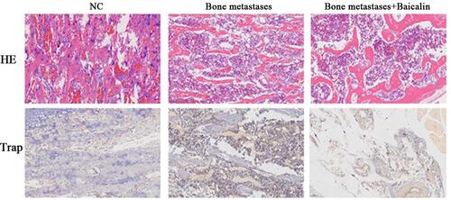
Figure 3 Baicalin inhibited the expression of iNOS in the Bone metastases cancer. iNOS was reported to be related with tumor metastasis. Fluorescein isothiocyanate (FITC) was labeled and showed bright yellow green fluorescence under fluorescence microscope. Immunofluorescence detection revealed that the expression of iNOS was significantly increased in the Bone metastases group and decreased after Bone metastases + Baicalin.
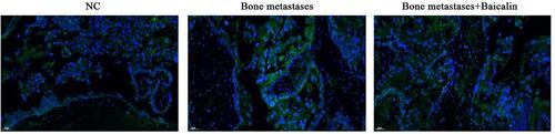
Figure 4 Baicalin inhibited the protein level of inflammatory factors (IL-1β, IL-6, TNF-α and PGE2) in the Bone metastases cancer shown by ELISA. Data were shown as mean ± SD. **p < 0.01, ***p < 0.001 was obtained from the NC group vs the Bone metastases group, the NC group vs the Bone metastases + Baicalin group, the Bone metastases group vs the Bone metastases + Baicalin group.
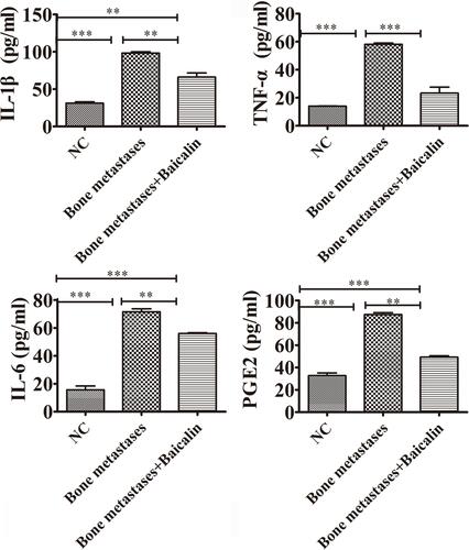
Table 1 Top 10 Enriched GO Pathways of DEGs Between the Bone Metastases Group and the NC Group
Table 2 Top 10 Enriched KEGG Pathways of DEGs Between the Bone Metastases Group and the NC Group
Table 3 Top 10 Enriched GO Pathways of DEGs Between the Baicalin Treatment Group and the NC Group
Table 4 Top 10 Enriched KEGG Pathways of DEGs Between the Baicalin Treatment Group and the NC Group
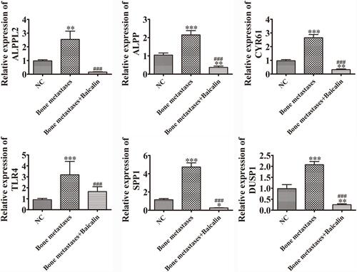
Figure 5 RT-qPCR results indicated that the mRNA expression level of ALPP, DUSP1, CYR61, ALPPL2, SPP1 and TLR4 were significantly up-regulated in the Bone metastases group compared with the NC group. By comparing the Bone metastases + Baicalin group and the NC group, the expression of these genes were significantly down-regulated. The RT-qPCR results of these genes were consistent with the results of the transcriptomic sequencing results. Data were shown as mean ± SD. *p < 0.05, **p < 0.01, ***p < 0.001 was obtained from the NC group vs the Bone metastases group or the Bone metastases + Baicalin group. ###p < 0.001 was obtained from the Bone metastases group vs the Bone metastases + Baicalin group.