Figures & data
Figure 1 The general function and mechanism of LncRNA. (1) In the nucleus, lncRNAs can guide chromatin modifiers and various transcriptional regulators to DNA, thereby inhibiting and/or activating gene expression. LncRNAs can be used as enhancers of target gene activation. They can also act as molecular baits to move proteins away from specific DNA locations. (2) In the cytoplasm, lncRNAs can be used as scaffolds to bring two or more proteins into the complex. In addition, they also act as sponges for other transcripts or proteins, as protein templates, or regulate mRNA degradation and translation.
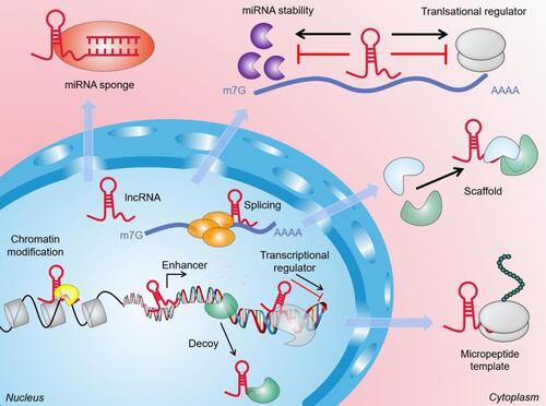
Table 1 LncRNAs are Involved in Regulating Oral Submucosal Fibrosis
Figure 2 LncRNAs participate in the regulation of oral submucosal fibrosis through multiple signaling pathways. LINC00974 and GAS5-AS1 activate the type I TGF-βR and recruit the receptor-activated Smads, the R-Smads (Smad2/3), which are phosphorylated to form a complex with Smad4. The inhibitory Smads (I-Smads) negatively regulate the Smad activation by competing with Smad2 and Smad3 for binding to the type I TGF-βR. Feedback loops between transcription factors and H19 also regulate the TGF-β induced EMT. Furthermore, Inhibition of ADAMTS9-AS2 and overexpression of HOTTIP involved in the PI3K/Akt signalling pathway could promote fBMFs activation and subsequently induce oral submucosal fibrosis.
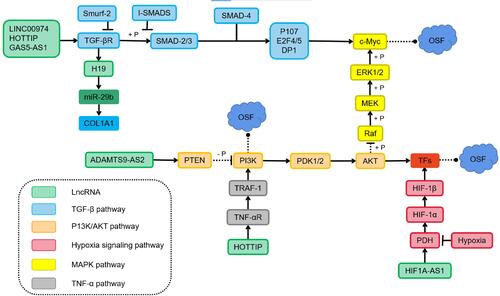
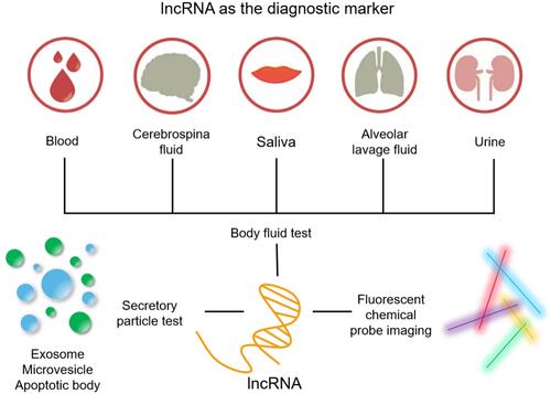
Figure 3 These images show various ways to use lncRNA as a diagnostic marker. Through minimally invasive sampling of cell-free fluids (blood, urine, saliva, cerebrospinal fluid, pleural effusion), quantitative detection of lncRNAs in exosomes, apoptotic bodies and extracellular vesicles. Fluorescent probe bioimaging technology can not only detect the abundance of clinically relevant biomarkers in OSF samples, but also detect their heterogeneity and spatial distribution.