Figures & data
Figure 1 Tabalumab binds membrane-bound BAFF on transfected cells or activated human monocytes.
Notes: (A) HEK293 cells were stably transfected with non-cleavable human BAFF. Membrane-bound BAFF was detected with Alexa 488-labeled tabalumab (gray histogram) compared with Alexa 488-labeled isotype control (black histogram). (B) Purified human CD14+ monocytes were activated with IFN-γ for 3 days. Membrane-bound BAFF was detected with Alexa 488-labeled tabalumab (gray histogram) compared with Alexa 488-labeled isotype control (black histogram). Data are from one experiment but representative of n=12 experiments (A) or ten donors (B).
Abbreviation: BAFF, B-cell activating factor.
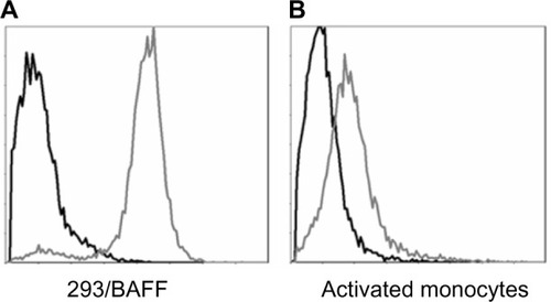
Figure 2 B-cells proliferate in response to soluble or membrane-bound BAFF.
Notes: (A) T1165.17 cells were stimulated with a dose titration of membrane-bound BAFF (■), soluble BAFF (○), or vector control cells (▲). Proliferation was measured using a colorimetric method for determining the number of viable cells at 44 hours. (B) Human CD19+ B-cells were co-stimulated with a dose titration of membrane-bound BAFF (■), soluble BAFF (○), or vector control cells (▲) in the presence of anti-IgM and IL-4. Proliferation was measured using 3H-thymidine incorporation at 72 hours. Data are presented as mean ± SD.
Abbreviations: BAFF, B-cell activating factor; CPM, counts per minute; Ig, immunoglobulin; IL, interleukin; SD, standard deviation.
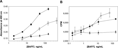
Figure 3 Tabalumab neutralization of soluble BAFF from multiple species.
Notes: IC50 was calculated for soluble human BAFF (IC50: 104 pM, 95% CI 96–112 pM), soluble cynomolgus monkey BAFF (IC50: 143 pM, 95% CI 126–162 pM), or soluble rabbit BAFF (IC50: 176 pM, 95% CI 170–182 pM). T1165.17 cells were stimulated with 365 pM BAFF and proliferation was measured using a colorimetric method for determining the number of viable cells at 44 hours. Data are presented as mean ± SD.
Abbreviations: BAFF, B-cell activating factor; CI, confidence interval; cyno, cynomolgus monkey; IC50, half maximal inhibitory concentration; SD, standard deviation.
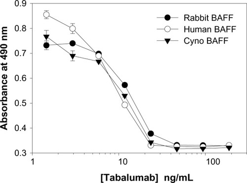
Figure 4 Tabalumab neutralizes soluble or membrane-bound human BAFF co-stimulation of B-cells.
Notes: (A) Primary human CD19+ B-cells were co-stimulated with 25 ng/mL soluble BAFF plus anti-IgM/IL-4 in the presence of a dose response of tabalumab or isotype control. Proliferation was measured using 3H-thymidine incorporation at day 5. (B) Primary human CD19+ B-cells were co-stimulated with 2.3 ng/mL soluble BAFF or 5.5 ng/mL membrane-bound BAFF plus anti-IgM/IL-4. Gray bars represent cells treated with 1 μg/mL tabalumab, black bars represent cells treated with 1 μg/mL isotype control. Stimulation with anti-IgM/IL-4 alone produced a relatively low proliferative signal of 3640 ± 592 CPM. Proliferation was measured using 3H-thymidine incorporation at day 3. Data are presented as mean ± SD.
Abbreviations: BAFF, B-cell activating factor; CPM, counts per minute; Ig, immunoglobulin; IL, interleukin; mem, membrane-bound; SD, standard deviation.
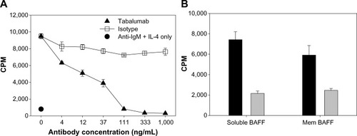
Figure 5 Tabalumab prevents BAFF binding to the three BAFF receptors.
Notes: HEK293 cells were transfected with either empty vector (A), human BR3 (B), human TACI (C), or human BCMA (D). Tabalumab or isotype control was pre-incubated with soluble biotinylated BAFF for 15 minutes prior to addition to each cell line. BAFF binding is visualized by addition of streptavidin-PE and the fluorescence is read on a flow cytometer. Black solid line represents cells incubated with buffer only; dark gray histogram represents cells and isotype control antibody + biotinylated BAFF; dotted line with open histogram represents cells and tabalumab + biotinylated BAFF.
Abbreviation: BAFF, B-cell activating factor; PE, phycoerythrin.
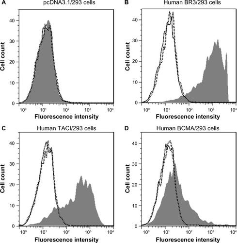
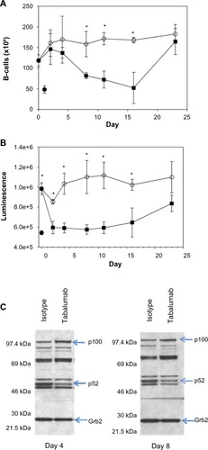
Figure 6 Administration of tabalumab to human BAFF transgenic mice leads to decreased B-cells and a reduction in non-canonical NF-κB signaling.
Notes: Tabalumab (■) or isotype control (○) was injected SC on day 0. Mice were sacrificed on days 2, 4, 8, 11, 16, and 23. (A) Splenic B-cells were enumerated by flow cytometry using B220; (B) p52 levels in the spleen were evaluated by a binding assay; (C) Western blot showing bands for p100 and p52 in splenocyte lysates; Grb2 was monitored to ensure equivalent loading of wells. * represents P-value <0.05. (•) represents an untreated non-transgenic mouse.
Abbreviations: BAFF, B-cell activating factor; SC, subcutaneously.