Figures & data
Figure 1 Luna 3D® (Benvenue Medical Inc., Santa Clara, CA, USA) lumbar interbody fusion cage.
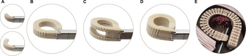
Figure 2 Axial view of Luna 3D® (Benvenue Medical Inc., Santa Clara, CA, USA) lumbar interbody fusion cage in situ – showing an ALIF cage-type footprint.
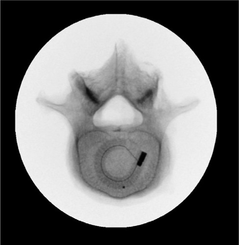
Figure 3 Lateral (A) and anteroposterior (B) radiographic views of initial deployment, middle component deployment (C and D), and fully deployed Luna 3D® (Benvenue Medical Inc., Santa Clara, CA, USA) lumbar interbody fusion cage (E and F).
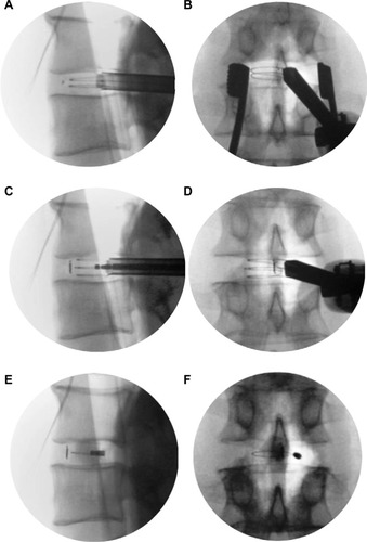
Table 1 Baseline patient characteristics (n=32)
Table 2 Operative data
Table 3 Clinical outcomes at 3 months (n=32)
Figure 4 Sagittal (A) and axial (B) MRI and sagittal CT (C) scans show grade I–II degenerative spondylolisthesis at L4–L5, total collapse of the disc, and severe spinal stenosis.
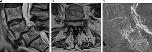
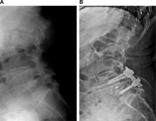
Figure 5 Radiographs showing preoperative grade I–II degenerative spondylolisthesis with total disc collapse at L4–5 (A) and stable construct with restoration of disc space 3 months after minimally invasive TLIF with Luna 3D® (Benvenue Medical Inc., Santa Clara, CA, USA) cage (B). At 3 months, the patient is doing well clinically and reported complete resolution of low back and lower extremity pain.