Figures & data
Figure 1 A schematic diagram depicting different brain regions and their involvement in specific fear processes (red), and more general role in cognitive/affective processing (blue).
Abbreviation: PTSD, posttraumatic stress disorder.
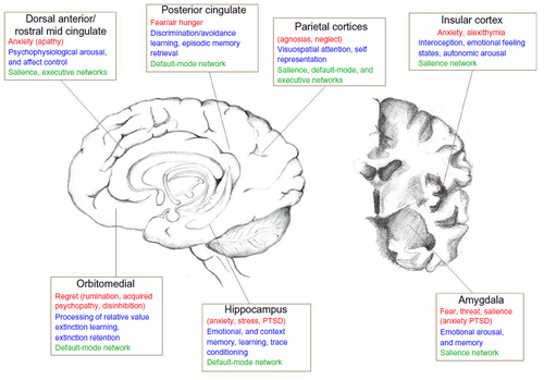
Figure 2 (A) Fear faces time-locked to distinct parts of the cardiac cycle were judged as more intense at systole on the heart beat relative to systole between heart beats. (B) Observing these fear faces induced a fear reaction in the brain that was also modulated by the cardiac cycle (eg, enhanced amygdala activation at systole), indicating that the contagion of fear responses is altered by bodily context. (C) Attentional capture of fear faces was exaggerated at systole, as demonstrated using the attentional blink paradigm to present fear faces at the cusp of conscious awareness.
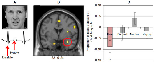
Figure 3 Brain and body interact to affect the perception and expression of fear.
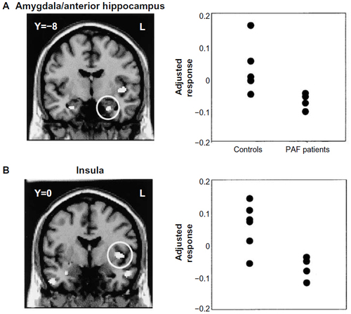
Figure 4 (A and B) Fear-conditioning and -extinction procedures can be used to demonstrate deficits in the retention of safety information over time in patients with PTSD relative to combat control participants.
Abbreviations: PTSD, posttraumatic stress disorder; CS, conditioned stimulus; CS+, CS with shock; CS-, without a shock.