Figures & data
Figure 1 Ginaton promoted the recovery of neurological function and reduced infarct volume after cerebral ischemia reperfusion in MCAO rats.
Notes: (A) mNSS scores of each group at 1 d, 7 d and 14 d after cerebral ischemia reperfusion; (B) time to walk across the whole beam in the beam-walking test; (C) representative images of infarct volume were shown by HE staining at 14 days after reperfusion; (D) quantitative evaluation of infarct volumes ratio in each group. Data were expressed as mean ± SEM. ***P<0.001 vs Sham group; #P<0.05, ###P<0.001 vs Model group; n=8–10.
Abbreviations: MCAO, middle cerebral artery occlusion; mNSS, modified neurological severity scores; HE, hematoxylin-eosin; SEM, standard error of mean.
Abbreviations: MCAO, middle cerebral artery occlusion; mNSS, modified neurological severity scores; HE, hematoxylin-eosin; SEM, standard error of mean.
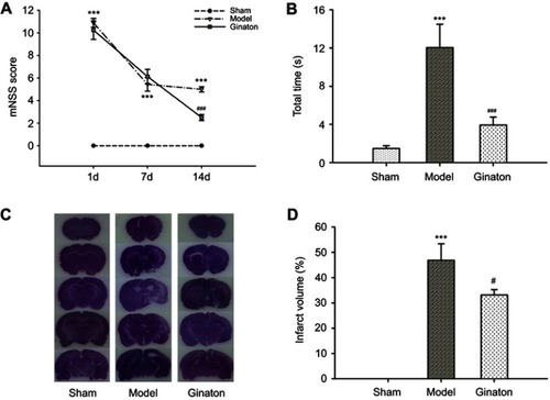
Figure 2 Ginaton reduced loss of NeuN-positive neurons in ischemic cortex penumbra after MCAO in rats.
Notes: (A) Representative immunohistochemical images of NeuN labeling in ischemic cortex penumbra and contralateral cortex in MCAO rats (magnification 200×, scale bar 50 μm); (B) quantification of NeuN-positive cells in contralateral cortex in different groups; (C) quantification of NeuN-positive cells in ischemic cortex in different groups. Data were expressed as mean ± SEM. *P<0.05 vs Sham group; #P<0.05 vs Model group; n=3.
Abbreviations: MCAO, middle cerebral artery occlusion; SEM, standard error of mean.
Abbreviations: MCAO, middle cerebral artery occlusion; SEM, standard error of mean.
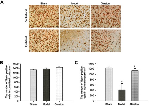
Figure 3 Ginaton induced autophagy via AMPK pathway in ischemic cortex of MCAO rats.
Notes: (A) Representative images and relative band intensity ratio of Beclin1 and LC3; (B) representative images and relative band intensity ratio of p-AMPKα, AMPKα, mTOR, and ULK1. The intensity of each band was normalized to that of β-actin. Data were expressed as mean ± SEM. *P<0.05, **P<0.01, ***P<0.001 vs Sham group; #P<0.05, ##P<0.01 ###P<0.001 vs Model group; n=3.
Abbreviations: AMPK, adenosine 5’-monophosphate (AMP)-activated protein kinase; MCAO, middle cerebral artery occlusion; LC3, light chain 3; mTOR, mammalian target of rapamycin; ULK1, unc-51-like kinase 1; SEM, standard error of mean.
Abbreviations: AMPK, adenosine 5’-monophosphate (AMP)-activated protein kinase; MCAO, middle cerebral artery occlusion; LC3, light chain 3; mTOR, mammalian target of rapamycin; ULK1, unc-51-like kinase 1; SEM, standard error of mean.
Figure 4 Ginaton maintained mitophagy and mitochondrial dynamics in ischemic cortex of MCAO rats.
Notes: (A) Representative images of immunoblots of Parkin, DRP1, OPA1 and β-actin; (B) relative band intensity ratio of Parkin/β-actin; (C) relative band intensity ratio of DRP1/β-actin; (D) relative band intensity ratio of OPA1/β-actin. The intensity of each band was normalized to that of β-actin. Data were expressed as mean ± SEM. **P<0.01, ***P<0.001 vs Sham group; #P<0.05, ###P<0.001 vs Model group; n=3.
Abbreviations: MCAO, middle cerebral artery occlusion; DRP1, dynamin-related protein 1; OPA1, optic atrophy 1; SEM, standard error of mean.
Abbreviations: MCAO, middle cerebral artery occlusion; DRP1, dynamin-related protein 1; OPA1, optic atrophy 1; SEM, standard error of mean.
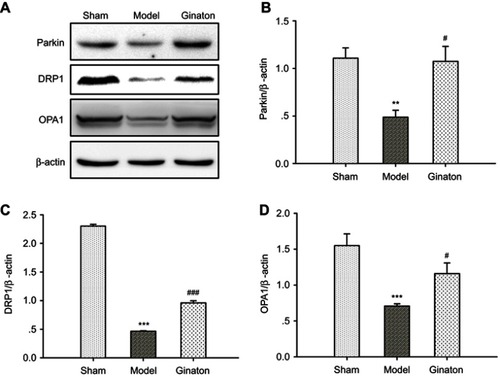
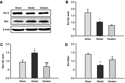
Figure 5 Ginaton inhibited apoptosis in ischemic cortex induced by MCAO in rats.
Notes: (A) Representative images of immunoblots of Bcl-2, Bax, and β-actin; (B) relative band intensity ratio of Bcl-2/β-actin; (C) relative band intensity ratio of Bax/β-actin; (D) relative band intensity ratio of Bcl-2/Bax. The intensity of each band was normalized to that of β-actin. Data were expressed as mean ± SEM. *P<0.05, **P<0.01 vs Sham group #P<0.05, ###P<0.001 vs Model group; n=3.
Abbreviations: MCAO, middle cerebral artery occlusion; SEM, standard error of mean.
Abbreviations: MCAO, middle cerebral artery occlusion; SEM, standard error of mean.