Figures & data
Table 1 Patient Demographics, Symptom Onset, Time Before TPK After Admission, Pre- and Postoperative Outcome
Figure 1 Total examinations performed and the number of positive examinations obtained for fungal infection in absolute numbers.
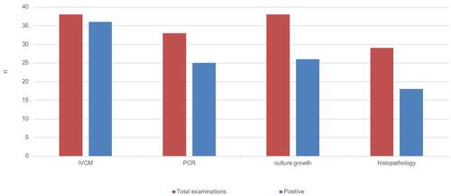
Figure 2 Slit lamp examination: 52-year-old female patient (case no. 15) with a Fusarium keratitis, referred 77 days after onset: (left) first presentation with CDVA of light perception only; (right) two years after receiving a second optical keratoplasty with a 7.6 mm graft due to endothelial graft decompensation after initial therapeutic keratoplasty with 13.0 mm graft; CDVA 1.0 LogMAR.
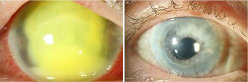
Figure 3 (top left) Slit lamp examination: a 44-year-old female patient (case no. 3) referred 12 days after symptom onset with CDVA hand movement only; (top right) follow-up examination 2 years after TPK, CDVA 0.1 LogMAR; (bottom left) a 36-year-old female patient (case no. 9) referred 4 days after symptom onset with CDVA 1.8 LogMAR; (bottom right) follow-up examination 1 year after TPK, CDVA 0.1 LogMAR.
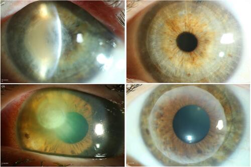
Table 2 Comparison of the Data of European Studies with Comparable Climate and Study Design
Figure 4 In vivo confocal microscopy findings. Many hyphae in the form of bright branched hyperreflective lines of varying width (left, middle, right) some of which appeared parallel (“railways tracks” - white arrow) in the corneal stroma, in the sense of fungal hyphae in context of fungal keratitis; (left) 44-year-old female patient as described in (case no. 3); (middle) 52-year-old female patient as described in (case no. 15); (right) 33-year-old female patient (case no. 21) with fungal hyphae at a depth over 200 µm in the cornea identified with in vivo confocal microscopy.
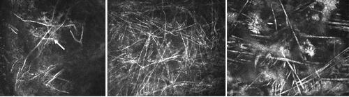
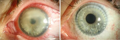
Figure 5 (left) A 20-year-old female patient at first presentation at our Department 17 days after symptom onset with CDVA hand movement only; (right) follow-up examination 2 months after initial TPK with 11.0 mm graft; CDVA 0.2 LogMAR.